The Great London:
Forensics

  • France: 6,000-yr-old skeletons in French pit were victims of violence

    France: 6,000-yr-old skeletons in French pit were victims of violence

    A gruesome discovery in eastern France casts new light on violent conflicts that took lives — and sometimes just limbs — around 6,000 years ago.

    6,000-yr-old skeletons in French pit were victims of violence
    A circular pit excavated in France (left) contains the remains of eight people probably 
    killed in a violent attack around 6,000 years ago. Seven severed left arms lay at the
     bottom of the pit. A diagram of the pit discoveries denotes bones of each individual
     in different colors [Credit: F. Chenal et al/Antiquity 2015, 
    © Bertrand Perrin/Antea]

    Excavations of a 2-meter-deep circular pit in Bergheim revealed seven human skeletons plus a skull section from an infant strewn atop the remains of seven human arms, say anthropologist Fanny Chenal of Antea Archéologie in Habsheim, France, and her colleagues.

    Two men, one woman and four children were killed, probably in a raid or other violent encounter, the researchers report in the December Antiquity. Their bodies were piled in a pit that already contained a collection of left arms hacked off by axes or other sharp implements. Scattered hand bones at the bottom of the pit suggest that hands from the severed limbs had been deliberately cut into pieces.

    It’s unclear who the arms belonged to. All the Bergheim skeletons have both their arms except for a man with skull damage caused by violent blows. His skeleton lacks a left arm, the researchers say. They have been unable to determine whether that arm ended up in the pit.

    Chenal’s group doesn’t know whether attackers targeted victims’ left arms for a particular reason. The arms could have been taken as war trophies, the team speculates.

    Radiocarbon dating of two bones indicates that individuals in the Bergheim pit lived roughly 6,000 years ago. From 6,500 to 5,500 years ago, during what’s known as the Neolithic period, one of the many ways of disposing of the dead in farming communities throughout Central and Western Europe was in circular pits.

    6,000-yr-old skeletons in French pit were victims of violence
    Fractures and stone-tool incisions appear on left forearm bones from 
    severed limbs found in a circular pit dating to 6,000 years ago 
    [Credit: F. Chenal et al/Antiquity 2015]

    Discoveries of human and nonhuman bones, as well as pottery, in these pits go back more than a century. The Bergheim pit provides the first evidence that people killed and mutilated in raids or battles were sometimes buried in circular pits, too, says study coauthor Bruno Boulestin, an anthropologist at the University of Bordeaux in France.

    Unusual deposits in Neolithic circular pits, such as attack victims and severed limbs at Bergheim, “may have been more common than previously expected,” says biological anthropologist Silvia Bello of the Natural History Museum in London, who did not participate in the new study. She suspects, for instance, that closer inspection of human bones previously found in circular pits elsewhere in Europe will reveal additional instances of violent deaths from a time when armed conflicts occurred between some communities.

    Bergheim’s brutalized victims spice up attempts to make sense of Neolithic circular pits. Many researchers regard these pits as remnants of storage silos that were put to other uses, possibly as receptacles for the bodies of people deemed unworthy of formal burials.

    Others argue that a large proportion of pits were dug as graves for high-ranking individuals, whose servants or relatives were killed to accompany them. Or, slaves might have been killed and put in pits as displays of wealth or as sacrifices to gods.

    Of 60 circular pits excavated in Bergheim in 2012 in advance of a construction project, 14 contained human bones. The researchers found skeletons or isolated bones of at least one to five individuals in each of 13 pits. The final pit contained the bodies and limbs described in the new paper.

    Joints of severed arms and skeletons in that pit were well-preserved, indicating that all had been placed there at or around the same time with a minimum amount of jostling disturbance. The pit also contained remains of a piece of jewelry made with a mussel’s valve, a stone arrowhead, a fragment of a pig’s jaw and two hare skeletons. The skeleton of a woman who had been put in the pit later lay on top of a sediment layer encasing those finds.

    Neither that woman nor human remains in the other Bergheim pits showed signs of violent death or limb loss.

    Author: Bruce Bower | Source: Science News [December 12, 2015]

  • Forensics: Five things you can learn from a Roman skeleton

    Forensics: Five things you can learn from a Roman skeleton

    The stories of Roman lives are written their bones: diet, disease, childbirth and trauma all leave their mark. Individual skeletons can tell rich tales, but the fullest information comes from large groups, when we can look at populations. So what can we learn about about a Roman community from their skeletons?

    Five things you can learn from a Roman skeleton
    The stories of Roman lives are written their bones: Roman skeleton found on
     at York University campus [Credit: University of York/PA]

    Whether they were a slave

    Slavery was ubiquitous in the Roman world, and some of its agonies are preserved on skeletons: those working in and living near Roman mines in Jordan were exposed to lead and copper at levels that would have been toxic, and caused a range of illnesses. The remains of people who were likely to have been slaves have also been found still wearing iron shackles, for instance in a subterranean room of a villa in Pompeii, and near the silver mines of Laurion in Roman-era Greece.

    Whether they played sports

    Among the human remains from ancient Herculaneum, which was destroyed by the eruption of Vesuvius at the same time as Pompeii, were a possible boxer, with typical fractures to his hands and nose, and a javelin-thrower whose bones reveal the same elbow problems experienced by modern athletes.

    How they died, who they loved

    At Dura-Europos in Syria, remains of Roman and Sasanian troops trapped in a siege mine beneath the walls of the ancient city reveal the brutal and violent reality of ancient conflict, including gas warfare. In the nearby cemetery, families were buried together in underground tombs, with women and children placed together.

    Five things you can learn from a Roman skeleton
    A well preserved Roman skeleton from the 2nd-4th century, found in a lead coffin 
    near Aldborough, North Yorkshire [Credit: Christopher Thomond/Guardian]

    Where people came from

    Even places like Roman Britain were diverse. Scientific methods (such as isotope analysis), as well as the study of graves and grave goods (the objects buried with a body) can tell us where a person was likely to have come from, or where they had links to. For instance, work on the cemeteries of Roman York has shown that people buried there came from other places in Britain, and much further afield in Europe, the Middle East and North Africa.

    The extent of childhood illnesses

    In the Roman world, children often didn’t make it to adulthood. Roman cemeteries such as Poundbury in Dorset include many children with rickets, scurvy and anaemia – survival rates were staggeringly low by modern Western standards. Infant and early childhood mortality was high in the Roman period, with 45% of children unlikely to survive past five years of age.

    So we can learn a lot about how a Roman may have lived from her or his remains, but, while skeletons are biological, bodies are cultured and contextual; they can be modified to fit ideals of beauty, status, or gender. Ultimately, Roman skeletons tell us that culture is a significant factor in determining difference: underneath it all, we’re pretty much the same collection of 206 interlocking parts.

    Dr Jen Baird and Dr Tim Reynolds from the Department of History, Classics and Archaeology, Birkbeck College, University of London, will be talking in depth about Roman skeletons at a Guardian Live/Birkbeck event on 21 November.

    Authors: Dr Jen Baird and Dr Tim Reynolds | Source: The Guardian [November 14, 2015]

  • Forensics: Intricate animal and flower tattoos found on Egyptian mummy

    Forensics: Intricate animal and flower tattoos found on Egyptian mummy

    A mummy from ancient Egypt was heavily tattooed with sacred symbols, which may have served to advertise and enhance the religious powers of the woman who received them more than 3,000 years ago.

    Intricate animal and flower tattoos found on Egyptian mummy
    The mummy's tattoos include two seated baboons depicted between a wadjet eye (top row), a symbol of protection 
    [Credit: Anne Austin]

    The newly reported tattoos are the first on a mummy from dynastic Egypt to show actual objects, among them lotus blossoms on the mummy’s hips, cows on her arm and baboons on her neck. Just a few other ancient Egyptian mummies sport tattoos, and those are merely patterns of dots or dashes.

    Especially prominent among the new tattoos are so-called wadjet eyes: possible symbols of protection against evil that adorn the mummy’s neck, shoulders and back.

    “Any angle that you look at this woman, you see a pair of divine eyes looking back at you,” says bioarchaeologist Anne Austin of Stanford University in California, who presented the findings last month at a meeting of the >American Association of Physical Anthropologists.

    Intricate animal and flower tattoos found on Egyptian mummy
    The mummy, found in the ancient village of Deir el-Medina, dates from 1300 to 1070 BC 
    [Credit: Anne Austin]

    Austin noticed the tattoos while examining mummies for the French Institute of Oriental Archaeology, which conducts research at Deir el-Medina, a village once home to the ancient artisans who worked on tombs in the nearby Valley of the Kings. Looking at a headless, armless torso dating from 1300 to 1070 bc, Austin noticed markings on the neck. At first, she thought that they had been painted on, but she soon realized that they were tattoos.

    Hidden history

    Austin knew of tattoos discovered on other mummies using infrared imaging, which peers more deeply into the skin than visible-light imaging. With help from infrared lighting and an infrared sensor, Austin determined that the Deir el-Medina mummy boasts more than 30 tattoos, including some on skin so darkened by the resins used in mummification that they were invisible to the eye. Austin and Cédric Gobeil, director of the French mission at Deir el-Medina, digitally stretched the images to counter distortion from the mummy’s shrunken skin.

    Intricate animal and flower tattoos found on Egyptian mummy
    Because the mummy's skin is distorted and covered in resin, it is difficult to see many tattoos — such as these 
    Hathor cows — with the naked eye [Credit: Anne Austin]

    The tattoos identified so far carry powerful religious significance. Many, such as the cows, are associated with the goddess Hathor, one of the most prominent deities in ancient Egypt. The symbols on the throat and arms may have been intended to give the woman a jolt of magical power as she sang or played music during rituals for Hathor.

    The tattoos may also be a public expression of the woman’s piety, says Emily Teeter, an Egyptologist at the University of Chicago’s Oriental Institute in Illinois. “We didn’t know about this sort of expression before,” Teeter says, adding that she and other Egyptologists were “dumbfounded” when they heard of the finding.

    Some tattoos are more faded than others, so perhaps some were made at different times. This could suggest that the woman’s religious status grew with age, Austin says.

    Intricate animal and flower tattoos found on Egyptian mummy
    Anthropologist Ghada Darwish Al-Khafif uses infrared imaging to examine tattoos on the mummy's back 
    [Credit: Anne Austin]

    Penetrating gaze

    She has already found three more tattooed mummies at Deir el-Medina, and hopes that modern techniques will uncover more elsewhere.

    Even infrared imaging can’t penetrate an intact mummy’s linen binding. But a nineteenth-century penchant for unwrapping mummies could enable the discovery of more tattoos, says Marie Vandenbeusch, a curator at the British Museum in London. Such examples could provide needed evidence “to really pinpoint the use of those tattoos”, she says.

    Austin argues that the scale of the designs, many of them in places out of the woman’s reach, implies that they were more than simple adornment.

    Intricate animal and flower tattoos found on Egyptian mummy
    This enhanced image of the mummy's skin reveals tattoos of two cows 
    [Credit: Anne Austin]

    The application of the tattoos “would’ve been very time consuming, and in some areas of the body, extremely painful”, Austin says. That the woman subjected herself to the needle so often shows “not only her belief in their importance, but others around her as well”.

    Author: Traci Watson | Source: Nature [doi:10.1038/nature.2016.19864] [May 09, 2016]

  • Genetics: Scientists sequence ancient British 'gladiator' genomes from Roman York

    Genetics: Scientists sequence ancient British 'gladiator' genomes from Roman York

    Cutting-edge genome technology in Trinity College Dublin has cast more light on a mystery that has perplexed archaeologists for more than a decade. The origins of a set of Roman-age decapitated bodies, found by York Archaeological Trust at Driffield Terrace in the city, have been explored, revealing a Middle Eastern body alongside native British.

    Scientists sequence ancient British 'gladiator' genomes from Roman York
    One of the skeletons excavated by York Archaeological Trust at Driffield Terrace
    [Credit: York Archaeological Trust]

    Archaeologists have speculated that the skeletons belonged to gladiators, although they could also have been soldiers or criminals. Several suffered perimortem decapitation and were all of a similar age – under 45 years old. Their skulls were buried with the body, although not positioned consistently – some were on the chest, some within the legs, and others at the feet.

    Although examining the skeletons revealed much about the life they lived – including childhood deprivation and injuries consistent with battle trauma – it was not until genomic analysis by a team from Trinity College Dublin, led by Professor of Population Genetics, Dan Bradley, that archaeologists could start to piece together the origins of the men.

    The Trinity College team recently published the first prehistoric Irish genomes and this analysis by Trinity PhD Researcher, Rui Martiniano, also breaks new ground as it represents the first genome analysis of ancient Britons.

    From the skeletons of more than 80 individuals, Dr Gundula Muldner of the University of Reading, Dr Janet Montgomery of the University of Durham and Malin Holst and Anwen Caffel of York Osteoarchaeology selected seven for whole genome analyses. Despite variation in isotope levels which suggested some of the 80 individuals lived their early lives outside Britain, most of those sampled had genomes similar to an earlier Iron Age woman from Melton, East Yorkshire. The poor childhood health of these men suggests that they were locals who endured childhood stress, but their robust skeletons and healed trauma, suggest that they were used to wielding weapons.

    Scientists sequence ancient British 'gladiator' genomes from Roman York
    The Roman-age skeletons from Driffield Terrace laid out in York's Guildhall 
    [Credit: York Archaeological Trust]

    The nearest modern descendants of the Roman British men sampled live not in Yorkshire, but in Wales. A man from a Christian Anglo-Saxon cemetery in the village of Norton, Teesside, has genes more closely aligned to modern East Anglia and Dutch individuals and highlights the impact of later migrations upon the genetic makeup of the earlier Roman British inhabitants.

    However, one of the decapitated Romans had a very different story, of Middle Eastern origin he grew up in the region of modern day Palestine, Jordan or Syria before migrating to this region and meeting his death in York.

    "Archaeology and osteoarchaeology can tell us a certain amount about the skeletons, but this new genomic and isotopic research can not only tell us about the body we see, but about its origins, and that is a huge step forward in understanding populations, migration patterns and how people moved around the ancient world," says Christine McDonnell, Head of Curatorial and Archive Services for York Archaeological Trust.

    "This hugely exciting, pioneering work will become the new standard for understanding the origins of skeletons in the future, and as the field grows, and costs of undertaking this kind of investigation fall, we may be able to refine our knowledge of exactly where the bodies were born to a much smaller region. That is a remarkable advance."

    Scientists sequence ancient British 'gladiator' genomes from Roman York
    The Roman skeletons were found at Driffield Terrace in York with their skulls placed between their legs,
     at their feet or on their chests [Credit: York Archaeological Trust]

    As well as Trinity College Dublin, the multi-disciplinary scientific analysis involved scientists from the University of York and The York Archaeological Trust, as well as the universities of Durham, Reading and Sheffield, University College London and the University Medical Centre in Utrecht. The research also included experts from York Osteoarchaeology Ltd, City of York Council and the Natural History Museum.

    The Roman skeletons sampled were all male, under 45 years old and most had evidence of decapitation. They were taller than average for Roman Britain and displayed evidence of significant trauma potentially related to interpersonal violence. All but one would have had brown eyes and black or brown hair but one had distinctive blue eyes and blond hair similar to the single Anglo-Saxon individual.

    The demographic profile of the York skeletons resembles the population structure in a Roman burial ground believed to be for gladiators at Ephesus. But the evidence could also fit with a military context—the Roman army had a minimum recruitment height and fallen soldiers would match the age profile of the York cemetery.

    Professor Dan Bradley, Trinity, said: "Whichever the identity of the enigmatic headless Romans from York, our sample of the genomes of seven of them, when combined with isotopic evidence, indicate six to be of British origin and one to have origins in the Middle East. It confirms the cosmopolitan character of the Roman Empire even at its most northerly extent."

    PhD Researcher and lead author, Rui Martiniano, Trinity, said: "This is the first refined genomic evidence for far-reaching ancient mobility and also the first snapshot of British genomes in the early centuries AD, indicating continuity with an Iron Age sample before the migrations of the Anglo-Saxon period."

    Professor Matthew Collins, of the BioArCh research facility in the Department of Archaeology at York, who co-ordinated the report on the research, "These genomes give the first snapshot of British genomes in the early centuries AD, showing continuity with the earlier Iron Age and evidence of migrations in the Anglo-Saxon period."

    The paper is published in >Nature Communications.

    Source: Trinity College Dublin [January 20, 2016]

  • Forensics: Slavery carried bilharzia parasites from West Africa to the Caribbean

    Forensics: Slavery carried bilharzia parasites from West Africa to the Caribbean

    Scientists used the full DNA sequences of Schistosoma mansoni parasites from Africa and the French Caribbean to discover the fluke's origins, map its historic transmission and identify the secrets of its success. Their findings, published in Scientific Reports, show how the global slave trade transported the disease from Senegal and Cameroon to Guadeloupe. Further genomic comparison with a closely related schistosome species that infects rodents reveals how the parasite has adapted to infecting human beings.

    Slavery carried bilharzia parasites from West Africa to the Caribbean
    The bilharzia-causing parasite, Schistosoma mansoni,  first infected humans as they fished
     in lakes in East Africa and was spread, first to West Africa and then to the New World, 
    by slave traders in 16th-19th Centuries, genomics reveals 
    [Credit: Wellcome Trust Sanger Institute]

    Schistosoma mansoni is a blood fluke (flatworm) that infects more than 250 million people worldwide and causes more than 11,000 deaths each year. Six years ago the Sanger Institute published the parasite's first full DNA sequence (genome); this latest study used that 'genetic map' to construct and compare the genomes of S. mansoni parasites gathered from across Africa and the New World, the majority of which were held at the Schistosomiasis Collection in the Natural History Museum, London.

    By analysing the differences between the human-infecting S. mansoni and its close relative, the rodent-infecting S. rodhaini, the scientists calculated that the two species evolved from a common ancestor approximately 107,000 to 148,000 years ago in East Africa. This finding suggests that the species is much 'younger' than previously thought.

    "The timing of the separation of the two species coincidences with the first archaeological evidence of fishing in Africa," explains Thomas Crellen, first author of the study from Imperial College London, the Sanger Institute and the Royal Veterinary College London. "The parasite develops in freshwater and infects people by burrowing through their skin. The introduction of fishing would have meant that people spent more time in the water, greatly increasing their chances of being infected."

    Analysing the differences between genomes from different locations also revealed the darker side of human history.

    "Comparing the S. mansoni genomes suggests that flukes in West Africa split from their Caribbean counterparts at some point between 1117AD and 1742AD, which overlaps with the time of the 16th-19th Century Atlantic Slave Trade," says Professor Joanne Webster from Imperial College London and the Royal Veterinary College. "During this period more than 22,000 African people were transported from West Africa to Guadeloupe by French slave ships, and the fluke was carried with them."

    Comparing the genomes of S. mansoni with S. rodhaini also revealed the genetic variations that have been positively selected over time in the human-infecting fluke and have been "fixed" into its DNA. It is likely that these variations are the evolutionary adaptations that have occurred to enable the fluke to successfully tunnel into, and thrive within, human beings.

    "When we looked for the differences between human-infecting S. mansoni DNA and its rodent infecting cousin S. rodhaini, we found two important variations. We found that changes to two genes in S. mansoni's DNA -- VAL21 and an elastase gene -appear to be important in allowing the fluke to enter and live in humans," says Dr James Cotton, senior author of the study from the Sanger Institute. "VAL genes produce proteins that cause allergic responses, so it is possible that the variation in VAL21 helps the fluke to hide from our immune systems. The elastase gene helps the parasite to burrow in to the body, by breaking down elastin -- a major component of human skin."

    It is hoped that exploring the genetic makeup of the fluke it will be possible to discover more about the processes the parasite relies on to infect humans and offer new opportunities to develop preventive and therapeutic interventions.

    Source: Wellcome Trust Sanger Institute [February 17, 2016]

  • Genetics: Mummies from Hungary reveal TB's Roman lineage

    Genetics: Mummies from Hungary reveal TB's Roman lineage

    Bodies found in a 200 year-old Hungarian crypt have revealed the secrets of how tuberculosis (TB) took hold in 18th century Europe, according to a research team led by the University of Warwick.

    Mummies from Hungary reveal TB's Roman lineage
    One of the 265 mummies resting in cardboard boxes in the Hungarian 
    Natural History Museum in Budapest, Hungary
    [Credit: AP/Bela Szandelszky]

    A new study published in Nature Communications details how samples taken from naturally mummified bodies found in an 18th century crypt in the Dominican church of Vác in Hungary have yielded 14 tuberculosis genomes, suggesting that mixed infections were common when TB was at peak prevalence in Europe.

    The research team included collaborators from the Universities of Warwick and Birmingham, University College London, the Hebrew University in Jerusalem and the Hungarian Natural History Museum in Budapest. Lead author Professor Mark Pallen, from Warwick Medical School, said the discovery was significant for current and future infection control and diagnosis.

    Professor Pallen said: “Microbiological analyses of samples from contemporary TB patients usually report a single strain of tuberculosis per patient. By contrast, five of the eight bodies in our study yielded more than one type of tuberculosis – remarkably from one individual we obtained evidence of three distinct strains.”

    The team used a technique called “metagenomics” to identify TB DNA in the historical specimens—that is direct sequencing of DNA from samples without growing bacteria or deliberately fishing out TB DNA. This approach draws on the remarkable throughput and ease of use of modern DNA sequencing technologies.

    Gemma Kay, first author on the paper says: “Poignantly, we found evidence of an intimate link between strains from in a middle-aged mother and her grown-up daughter, suggesting both family members died from this devastating infection.”

    The team used the 18th century sequences to date the origin of the lineage of TB strains commonly found in Europe and America to the late Roman period, which fits in with the recent controversial suggestion that the most recent common ancestor of all TB strains occurred as recently as six thousand years ago.

    Professor Pallen said: “By showing that historical strains can be accurately mapped to contemporary lineages, we have ruled out, for early modern Europe, the kind of scenario recently proposed for the Americas—that is wholesale replacement of one major lineage by another—and have confirmed the genotypic continuity of an infection that has ravaged the heart of Europe since prehistoric times.”

    Professor Pallen added that with TB resurgent in many parts of the world, the struggle to contain this ancient infection was far from over. He concludes: “We have shown that metagenomic approaches can document past infections. However, we have also recently shown that metagenomics can identify and characterize pathogens in contemporary samples, so such approaches might soon also inform current and future infectious disease diagnosis and control.”

    For more photos of the Hungarian mummies visit the website Morbid Anatomy.

    Source: University of Warwick [April 07, 2015]

  • Forensics: Palaeolithic remains show cannibalistic habits of human ancestors

    Forensics: Palaeolithic remains show cannibalistic habits of human ancestors

    Analysis of ancient cadavers recovered at a famous archaeological site confirm the existence of a sophisticated culture of butchering and carving human remains, according to a team of scientists from the Natural History Museum, University College London, and a number of Spanish universities.

    Palaeolithic remains show cannibalistic habits of human ancestors
    Human skull-cup uncovered in Gough's Cave, Somerset [Credit: The Trustees
     of the Natural History Museum, London]

    Gough's Cave in Somerset was thought to have given up all its secrets when excavations ended in 1992, yet research on human bones from the site has continued in the decades since.

    After its discovery in the 1880s, the site was developed as a show cave and largely emptied of sediment, at times with minimal archaeological supervision.

    The excavations uncovered intensively-processed human bones intermingled with abundant butchered large mammal remains and a diverse range of flint, bone, antler, and ivory artefacts.

    Palaeolithic remains show cannibalistic habits of human ancestors
    Fragments of human skull and mandibles from Gough's Cave [Credit: The Trustees 
    of the Natural History Museum, London]

    New radiocarbon techniques have revealed remains were deposited over a very short period of time, possibly during a series of seasonal occupations, about 14,700 years ago.

    Dr Silvia Bello, from the Natural History Museum's Department of Earth Sciences, lead researcher of the work said, "The human remains have been the subject of several studies. In a previous analysis, we could determine that the cranial remains had been carefully modified to make skull-cups. During this research, however, we've identified a far greater degree of human modification than recorded in earlier. We've found undoubting evidence for defleshing, disarticulation, human chewing, crushing of spongy bone, and the cracking of bones to extract marrow."

    Palaeolithic remains show cannibalistic habits of human ancestors
    Human chewing damage on a rib bone, showing breaks made by saw-teeth (white arrows), 
    dug out furrows (B), and slicing cut marks (C). Scale bar  = 250 μm. 
    [Credit: The Trustees of the Natural History Museum, London]

    The presence of human tooth marks on many of the bones provides incontrovertible evidence for cannibalism, the team found. In a wider context, the treatment of the human corpses and the manufacture and use of skull-cups at Gough's Cave has parallels with other ancient sites in central and western Europe.

    But the new evidence from Gough's Cave suggests that cannibalism during the 'Magdalenian period' was part of a customary mortuary practice that combined intensive processing and consumption of the bodies with the ritual use of skull-cups.

    Simon Parfitt, of University College London, said, "A recurring theme of this period is the remarkable rarity of burials and how commonly we find human remains mixed with occupation waste at many sites. Further analysis along the lines used to study Gough's Cave will help to establish whether the type of ritualistic cannibalism practiced there is a regional ('Creswellian') phenomenon, or a more widespread practice found throughout the Magdalenian world."

    Source: Natural History Museum [April 16, 2015]

  • Forensics: New research to shed fresh light on the impact of industrialisation

    Forensics: New research to shed fresh light on the impact of industrialisation

    The Museum of London today announced a ground-breaking research project to explore the effects of industrialisation on Londoners. The research hopes to uncover new clues about the very nature of disease and how it has affected people as Britain has moved into the age of industrialisation.

    New research to shed fresh light on the impact of industrialisation
    Some of London’s most important skeletons will be digitally x-rayed and scanned 
    in a new archaeological investigation linking “man-made” diseases with
     the industrialisation of the city [Credit: © Museum of London]

    The research has been made possible by a City of London Archaeological Trust grant from a bequest made by the late Rosemary Green.

    John Schofield, Secretary of the City of London Archaeological Trust, said: “The City of London Archaeological Trust is very happy that the Rosemary Green bequest is used to gather this cutting-edge data on the signs of industrialisation in the skeletal collections on the Museum of London.”

    Leading the project is Jelena Bekvalac, based at the Museum of London’s Centre for Human Bioarcheology, along with her research team, Gaynor Western and Mark Farmer.

    Jelena Bekvalac, said: “The most tangible evidence we have for the long-term consequences of the industrialisation process upon us is, quite simply, written in our bones. Using the very latest digital technology, we will examine the skeletal remains of over 1,000 adult men and women from industrial-era London in addition to a further 500 skeletons from the medieval metropolis.

    New research to shed fresh light on the impact of industrialisation
    More than 1,000 adult men and women from the industrial revolution period,
     as well as 500 more from the medieval metropolis, will be used in quest to
     find out how the era affected the population at the Museum of London’s 
    Centre for Human Bioarchaeology [Credit: © Museum of London]

    “Modern health trends have seen a shift towards increasing life expectancy but we want to look again at what are often thought of as ‘man-made’ conditions like obesity and cancer. Given today’s more sedentary lifestyles, far removed from the physically active and natural existence of most of our forebears, there are some big questions about the origins of these diseases and how they relate to the modern environment.”

    The research aims to address some of these questions by analysing diseases affecting the human skeleton. The museum will use the latest clinical techniques, including direct digital radiography, CT scanning and 3D modelling, to get a better understanding of what the bones tell us and to assess their change over time. The research aims to examine the influence of the industrial revolution, a pivotal catalyst in the formation of the modern age, on the changing nature of disease – from the medieval and post-medieval periods through to the present day.

    The project offers an exciting opportunity to digitise some of London’s most important skeletal collections, while simultaneously telling a new story about the health of Londoners over time.

    This work will culminate in the creation of an extensive new interactive digital resource that can be explored online. Jelena Bekvalac plans to make an immediate start on the digital scanning. She aims to publish her team’s findings as soon as possible and deliver a series of lectures about the work.

    Source: Museum of London [April 24, 2015]

  • Europe: Skeletal marker of physiological stress might indicate good, rather than poor, health

    Europe: Skeletal marker of physiological stress might indicate good, rather than poor, health

    Biological anthropologist Sharon DeWitte studies ancient skeletons that can open a window onto the human history she hopes to illuminate. But as she and graduate student Samantha Yaussy show in a recently published study, some of the markers on the skeletons that scientists use to decipher the past might need to be looked at in a new light.

    Skeletal marker of physiological stress might indicate good, rather than poor, health
    Horizontal grooves in skeletal teeth (linear enamel hypoplasia) are an indication of physiological stress on an
     individual in early childhood, between six months and six years of age [Credit: Sharon DeWitte]

    DeWitte, one of the world's foremost experts on the Black Death, which killed one-third to one-half of Europe's inhabitants over just seven years in a mid-1300s pandemic, is also interested in the periods before and after the Black Death, both in times of famine and, for the medieval era, relative plenty.

    "This was a time when you had all of these other stressors existing, like multiple infectious diseases, people living in really crowded conditions, lack of hygiene," DeWitte says. "I'm interested in looking at how famine might have affected subsequent patterns of health and risks of mortality."

    In research recently published in the >American Journal of Physical Anthropology, Yaussy and DeWitte worked with skeletal remains excavated from London's St. Mary Spital cemetery, which was in use from about 1120 to 1540 A.D. The cemetery was organized well enough over those years that modern-day researchers can assign burial groupings according to location to a number of shorter time periods within that larger 420-year time frame. Because they were focused on the effects of famine, the scientists specifically excluded burials from the Black Death era in the study.

    The way that people were buried in plots also provided important data. Large group burials within a single grave were taken to be the result of catastrophic famine losses over short periods of time, whereas single interments were categorized as "attritional" deaths, that is, deaths in "normal" times.

    Using a total of more than 1,500 individual adult remains that were almost evenly divided between famine deaths and normal deaths, DeWitte and Yaussy carefully examined the skeletons for markers that are indicative of stress during the individual's lifetime.

    One stress marker they looked for is called "linear enamel hypoplasia" (LEH), which is a horizontal groove on a tooth that results from childhood physiological stress or trauma sometime between 6 months and 6 years of age, when the tooth is forming in the jaw.

    "It could be the result of an infection, lack of nutrition, or even breaking a leg," says Yaussy. "Your enamel just stops creating itself for a day or a little longer. It starts back up again, but during that time when it was shut off, there's no enamel on that portion of the teeth, and it leaves a groove."

    They found that that stress marker, LEH, was significantly correlated with famine. "It was a pretty sensitive indicator of famine burials," Yaussy says. "Having that early life insult seems to instigate this pattern of lifelong frailty, so when a famine event occurs, it causes mortality in those individuals."

    Another indicator of life stress, though, turned out to be a horse of a different color. Using shin bones (tibia), the researchers looked for what's called periosteal lesions. It's a place on the bone where new growth on the surface has occurred in response to physical or physiological stress.

    "When it's put under stress, and it can be from something like an infection, or a break, or even just stress from carrying heavy buckets all day, bone can grow onto itself and strengthen itself," Yaussy says. "These are nonspecific -- we're not necessarily saying that it was an infection that caused it, or that it was from someone hitting their shin repeatedly. I just see that there was bone growth there, so there's some stressor that's causing the bone to generate more bone."

    In contrast to people with the LEH indicator, individuals with areas of bone regrowth were correlated with periods of normal, or attritional, death. That result came as a something of a surprise to colleagues in the field.

    "This project was a bit of a shock to some of the people at the paleopathology meetings where we presented it," Yaussy says. "A lot of people have been thinking that things like periosteal lesions are bad, that people who had them weren't especially healthy. But this study in part might be saying that those people were actually pretty healthy. It takes time, a couple of weeks, to build up this bone, so it could be that if there was some stress event that was substantial enough to kill a person before they could even register a response, we wouldn't be seeing it on the bone now."

    The unexpected result helps inform DeWitte in the larger goal of looking at what happens to populations after crises, such as the Black Death or famines, pass, and particularly how cultural norms might influence the outcomes.

    "What I'm interested in going forward is looking at access to resources before the Black Death, during normal conditions during famine conditions, and then after the Black Death, again during normal and famine conditions," Dewitte says. "Seeing if there were social factors that affected people's access to resources when there was little available and then when they became abundant again."

    "I think this might have implications for living populations, understanding how social, economic and political factors affect access to resources beyond the actual amount of resources that are available."

    Author: Steven Powell | Source: University of South Carolina [May 02, 2016]

  • Near East: Face of 9,500 year old Neolithic man from Jericho reconstructed

    Near East: Face of 9,500 year old Neolithic man from Jericho reconstructed

    Archaeologists from the British museum have reconstructed an ancient man's face, allowing visitors to see what he looked like for the first time.

    Face of 9,500 year old Neolithic man from Jericho reconstructed
    Archaeologists from the British Museum have reconstructed the face of a man who lived 9,500 
    years ago in the city of Jericho, now found in the Palestinian territories near the West Bank 
    [Credit: Copyright: The Trustees of the British Museum]

    The man lived 9,500 years ago in the holy city of Jericho, now found in the Palestinian territories near the West Bank.

    The 'Jericho skull' was found by British archaeologists in 1953, but until now nobody knew what the he had looked like.

    Scientists still don't know the man's true identity, but they speculate that he was once someone of great importance.

    This is based on the amount of care people had taken to fill his skull with plaster once he had died, almost 10,000 years ago.

    Face of 9,500 year old Neolithic man from Jericho reconstructed
    The Jericho Skull. Tell es-Sultan, Jericho, Palestinian Authority. Human bone, plaster, shell, soil. 
    About 8200-7500 BC, Middle Pre-pottery Neolithic B period 
    [Credit: Copyright: The Trustees of the British Museum]

    Back then, plastered skulls were a form of ritual burial, like the Egyptians' infamous mummification burials.

    The gruesome practice involved removing the corpse's skull and filling it with plaster, before painting over the dead person's face and filling his eye sockets with shells.

    These remains were likely put on display for locals while the rest of the body was buried under the family home.

    The Jericho skull was found nestled alongside several other plastered skulls, but was by far the most well-preserved of the group.


    'He was certainly a mature individual when he died, but we cannot say exactly why his skull, or for that matter the other skulls that were buried alongside him, were chosen to be plastered,' British Museum curator Alexandra Fletcher told >Seeker.

    'It may have been something these individuals achieved in life that led to them being remembered after death.'

    Before the reconstruction, the ancient skull showed few human features due to the plaster pasting over most of its features.

    To investigate the grim burial practice, the scientists sent the skull off for a scan at the Imaging and Analysis Centre at London's Natural History Museum.

    Face of 9,500 year old Neolithic man from Jericho reconstructed

    Face of 9,500 year old Neolithic man from Jericho reconstructed
    Through the CT scans, the team discovered that the ancient man was missing
    a jaw underneath the plaster, and had lines of decaying teeth 
    [Credit: Copyright: Trustees of the British Museum]

    Here, a complete micro-CT scan unveiled a ream of new information about the skull, and inspired the Museum to undertake a full plaster reconstruction.

    Through the CT scans, the team discovered that the ancient man was missing a jaw underneath the plaster, and had lines of decaying teeth.

    They could see he had suffered a broken nose at some point in his life.

    He had also undergone head-binding, a traditional practice in which the skull of a human being is deformed intentionally, usually by forcefully distorting a child's skull.

    Face of 9,500 year old Neolithic man from Jericho reconstructed

    Face of 9,500 year old Neolithic man from Jericho reconstructed
    The plaster skull during the reconstruction process showcasing the ancient man's muscle and tissue build
    [Credit: Copyright: The Trustees of the British Museum]

    'Head binding is something that many different peoples have undertaken in various forms around the world until very recently,' Fletcher told Seeker.

    'In this case, the bindings have made the top and back of the head broader - different from other practices that aim for an elongated shape. I think this was regarded as a 'good look' in Jericho at this time.'

    All of the newly gathered details allowed the team to make an accurate plaster reconstruction of the man's head.

    And while the fascinating new model provides fresh insight into the man's life, plenty more work needs to be done to discover more about his history and culture.

    Face of 9,500 year old Neolithic man from Jericho reconstructed
    Side and front views of the reconstruction. The effect of the head binding is just visible 
    [Credit: Copyright: Trustees of the British Museum]

    The team hopes to gather DNA samples from the skull in future, laying out 10,000 year-old genes for investigation.

    But the process would be risky - it's likely to damage the skull and useful results aren't guaranteed.

    'If we were able to extract DNA from the human remains beneath the plaster, there is currently a very slight chance that we would be able to find out this individual's hair and eye colour,' Fletcher said.

    'I say a slight chance because the DNA preservation in such ancient human remains can be too poor to obtain any information.'

    The reconstructed face will be on display at the British Museum in London from next Thursday until mid-February.

    Author: Harry Pettit | Source: Daily Mail Online [December 09, 2016]

  • UK: Ancient Britons' teeth reveal people were 'highly mobile' 4,000 years ago

    UK: Ancient Britons' teeth reveal people were 'highly mobile' 4,000 years ago

    Archaeologists have created a new database from the teeth of prehistoric humans found at ancient burial sites in Britain and Ireland that tell us a lot about their climate, their diet and even how far they may have travelled. In a paper, led by Dr Maura Pellegrini from the University of Oxford, researchers say that individuals in prehistoric Britain were highly mobile.

    Ancient Britons' teeth reveal people were 'highly mobile' 4,000 years ago
    Ancient Britons' teeth were analysed for clues as to where they had grown up 
    [Credit: Mandy Jay]

    The study is part of the international Beaker People project lead by Professor Mike Parker Pearson of University College London, and involves scientists from many institutions, including the universities of Oxford, Durham, Bradford, University College London, and the Max Planck Institute for Evolutionary Anthropology in Leipzig, Germany. The paper says most of the teeth in the collection date back to Chalcolithic and Early Bronze Age periods (from 2500 to 1500 BC) and the analysis, published in the journal >Scientific Reports, suggests not only were people moving around their own country but may also have travelled to and from continental Europe.

    Researchers describe how tests on tooth fragments using an oxygen isotope analysis of tooth enamel provides evidence of where an individual lived when the tooth formed. Oxygen, a naturally occurring element in the environment, is absorbed by plants and animals and fixed in the mineral component of mammalian teeth, with an isotopic composition related to the environment in which that individual spent their childhood.  Based on the theory that prehistoric people would have sourced water and food locally, the team were able to geographically map the oxygen isotopic variability in the landscape of Britain and Ireland thereby providing a guide to where individuals sampled had lived as children.

    Ancient Britons' teeth reveal people were 'highly mobile' 4,000 years ago
    Woodhenge, one of the locations the variability in the isotope values was found to be particularly 
    marked in individuals [Credit: WikiCommons]

    An analysis of the teeth of those buried in the Stonehenge region, the Peak District, and the hills of the Yorkshire Wolds (in East Riding and North Yorkshire) show many were not 'local' to their final resting place. They were drawn from far and wide, sometimes to focal points containing sacred monuments. The variability in the isotope values was found to be particularly marked in individuals recovered from Woodhenge, a timber circle situated near Stonehenge; Bee Low, a Bronze Age round cairn in the Peak District, and Garton Slack in Yorkshire where there is a complex range of barrow types and burial practices.

    Tooth enamel fragments from 261 individual teeth were tested with researchers focusing on the central part of the tooth crown in each case. The teeth sampled from these individuals mineralise from the age of two years up to 8 years old, providing the clues to the environmental conditions, including the water they drank as a child. The possibility that people were outsiders who came into areas where they eventually died was calculated by comparing their values with the 'isoscape' information gleaned from most of the other samples in each area, as it was assumed the latter represented 'local' individuals. As individuals' signatures in the teeth were matched with areas where the majority, or 'local' people, were found, the researchers identified those who had lived in other areas as children.

    Source: University of Oxford [October 08, 2016]

  • Fossils: Ear ossicles of modern humans and Neanderthals: Different shape, similar function

    Fossils: Ear ossicles of modern humans and Neanderthals: Different shape, similar function

    A research team led by scientists of the Max Planck Institute for Evolutionary Anthropology scanned the skulls of Neandertals and found the small middle ear ossicles, which are important for hearing, still preserved within the cavities of the ear. To their surprise, the Neandertal ossicles are morphologically distinct from the ossicles of modern humans. Despite the differences in morphology, the function of the middle ear is largely the same in the two human species.

    Ear ossicles of modern humans and Neanderthals: Different shape, similar function
    Tympanic membrane (grey), ossicular chain (yellow, green, red), and bony inner ear (blue) of a modern human 
    with a One-Eurocent coin for scale [Credit: © A. Stoessel & P. Gunz]

    The authors relate the morphological differences in the ossicles to different evolutionary trajectories in brain size increase and suggest that these findings might be indicative of consistent aspects of vocal communication in modern humans and Neandertals. These findings are also of importance for shedding light on the emergence of human spoken language, which can only be inferred indirectly from the archaeological and fossil record.

    The three bones of the middle ear (hammer, anvil, stapes) make up the ossicular chain. This bony chain, which is found in all mammals is dedicated to the transmission of sound waves from the tympanic membrane to the inner ear and helps in amplifying the energy of airborne sound in order to allow the sound wave to travel within the fluid-filled inner ear.

    Moreover, the ear ossicles are not only important for correct hearing but are also the smallest bones of our body. Thus, it does not surprise that the ossicles are among the most rarely found bones in the mammalian fossil record including the one of human ancestors. Given their important role in audition this lack of knowledge has ever been frustrating for researchers interested in studying hearing capacities of extinct species.

    Tiny bones still present

    This also applies to our closest extinct relatives - the Neandertals whose communicative capacities including existence of human spoken language is a major scientific debate ever since the first discovery of Neandertal remains. A research team led by Alexander Stoessel from the Max Planck Institute for Evolutionary Anthropology in Leipzig used high-resolution computer tomography scans of Neandertal skulls and systematically checked for ossicles that potentially became trapped within the cavity of the middle ear.

    And indeed, the researchers found ear ossicles in 14 Neandertal individuals coming from sites in France, Germany, Croatia and Israel, resulting in the largest sample of ear ossicles of any fossil human species. “We were really astonished how often the ear ossicles are actually present in these fossil remains, particularly when the ear became filled with sediments” says lead researcher Alexander Stoessel.

    After virtually reconstructing the bones, the team - which also included scientist from the Friedrich-Schiller University in Jena and the University College in London – compared them to ossicles of anatomically modern humans and also chimpanzees and gorillas which are our closest living relatives.

    Since ossicles are not only small but also complex-shaped the researchers compared them by means of three-dimensional analysis that uses a much larger number of measuring points allowing for examination of the three-dimensional shape of a structure. “Despite the close relationship between anatomically modern humans and Neandertals to our surprise the ear ossicles are very differently shaped between the two human species” says Romain David who was involved in the study.

    Based on the results of the morphological comparison the research team examined the potential reasons for these different morphologies. In order to see if these differences may affect hearing capacity of Neandertals and modern humans or reflects a tight relationship with the base of the skull they also analyzed the structures surrounding the ear ossicles. The outcome of this analysis was surprising, again since the functional parameters of the Neandertal and modern human middle ear are largely similar despite contrasting morphologies.

    Similar communication skills in archaic humans

    Instead, the team found the ear ossicles strongly related to the morphology of the surrounding cranial structures which also differ between the two human groups. The reseachers attribute these differences to different evolutionary trajectories that Neandertals and modern humans pursued in order to increase their brain volume which also impacted the structures of the cranial base which the middle ear is a part of.

    “For us these results could be indicative for consistent aspects of vocal communication in anatomically modern humans and Neandertals that were already present in their common ancestor” says Jean-Jacques Hublin who is an author of this study and continues “these findings should be a basis for continuing research on the nature of the spoken language in archaic hominins”.

    The findings are published in >Proceedings of the National Academy of Sciences.

    Source: Max Planck Institute for Evolutionary Anthropology [September 27, 2016]

  • UK: Two ancient Chinese skeletons found in London Roman cemetery

    UK: Two ancient Chinese skeletons found in London Roman cemetery

    Two ancient skeletons unearthed at a cemetery in London may have been of Chinese origin, overturning longstanding assumptions about the history of the Roman Empire and Britain's capital city.

    Two ancient Chinese skeletons found in London Roman cemetery
    Parts of a skeleton found at Lant Street [Credit: Museum of London]

    Using cutting-edge techniques, a team of archaeologists and scientists examined dental enamel samples from over 20 sets of human remains dated from between the 2nd and 4th centuries AD.

    Dr Rebecca Redfern, curator of human osteology at the Museum of London, revealed two of the skeletons found at the site in Lant Street, Southwark, had been identified as possibly being of Chinese origin.

    "This is absolutely phenomenal. This is the first time in Roman Britain we've identified people with Asian ancestry and only the 3rd or 4th in the empire as a whole", she told BBC Radio 4's The World at One,

    The find challenges the dominance of the traditional view that Roman Britain, and specifically “Londinium” as it was then known, was a relatively homogenous society.

    It also suggests the Roman and Chinese empires may have had more interaction than many historians had previously thought.

    Crucially though, it raises the possibility that trade took place between Rome and China outside of the famous Silk Road.

    Two ancient Chinese skeletons found in London Roman cemetery
    One of the skeletons found at the site in Lant Street, Southwark 
    [Credit: Museum of London]

    While previous archaeological work has shown the multicultural nature of the ancient city and its importance as a major trading hub, this is only the second time the bones of an individual of possibly Chinese origin have been found at a Roman site, the first being the discovery of a possibly Asian man in Vagnari, Italy.

    Writing in >The Journal of Archaeological Science, Dr Redfern said: “The expansion of the Roman Empire across most of western Europe and the Mediterranean, led to the assimilation and movement of many ethnically and geographically diverse communities.

    "Its power and wealth meant that it also had trade connections for raw materials and products, such as silk throughout Europe, Africa and also to the east, including India and China.

    "Many people travelled, often vast distances, for trade or because of their occupation, for example in the military, or their social status, for example if they were enslaved."

    Archaeologists and historians are divided as to the the explanation behind the possible presence of Chinese individuals in Roman Britain.

    The findings raise the possibility that Chinese traders settled in the area, and may have even set up their own trading communities.

    However, in her journal article, Dr Redfern went on to add: “It may well be that these individuals were themselves or were descended from enslaved people originating from Asia, as there were slave-trade connections between India and China, and India and Rome."

    Author: James Somper | Source: Independent [September 27, 2016]

  • Polynesia: Forensic analysis of pigtails to help identify original 'mutineers of H.M.S. Bounty'
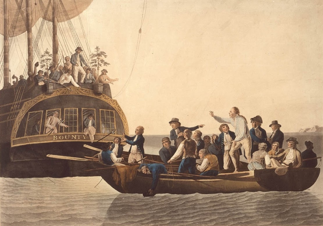
    Polynesia: Forensic analysis of pigtails to help identify original 'mutineers of H.M.S. Bounty'

    Ten pigtails of hair thought to be from seven mutineers of "Mutiny on the Bounty" fame and three of their female Polynesian companions will be analysed in a new collaboration between the Pitcairn Islands Study Centre at Pacific Union College (California, US) and the forensic DNA group at King's College London (UK).

    Forensic analysis of pigtails to help identify original 'mutineers of H.M.S. Bounty'
    A hand-coloured engraving dated 1790, depicting Captain William Bligh being 
    cast adrift from The Bounty [Credit: State Library of New South Wales]

    The forensic DNA group at King's has been sent hair strands from the ten pigtails, which are currently on display in the California-based centre, to help establish as much information as possible on their origins.

    As the pigtails purportedly date back to the pre-1800s, the King's team will first attempt to extract DNA from the historical hair samples after cleaning the outside and then digesting the hair matrix using a chemical process. Nuclear DNA is not found in hair shafts, only the roots which are not available here; however, mitochondrial DNA may be present. If sufficient mitochondrial DNA can be collected, the first step will be to investigate the ancestral origins of the owners of the pigtails.

    Unlike nuclear DNA, mitochondrial DNA does not discriminate between all individuals as people sharing a common maternal ancestor will also share a similar profile. However, this type of DNA can provide some indication of maternal geographic origin e.g. whether someone is likely to be of European descent, so the team will aim to establish whether the hair samples do indeed come from seven Europeans and three Polynesian individuals, as the documentation accompanying the samples suggests.

    Further, more detailed identification will require genealogical methods to trace the ancestors of the pigtail owners, to be able to link samples to names from historical records and other sources of information. A lot has been written about the possible descendants of the mutineers but this information will not be helpful with regards to the male mutineers; instead, their maternal line will need be traced. The study will therefore try to identify their maternal ancestors, such as their respective mothers and maternal grandmothers, and research other direct female descendants down to individuals living today.

    Forensic analysis of pigtails to help identify original 'mutineers of H.M.S. Bounty'
    Hair from the collection of pigtails donated to the Pitcairn Islands Study Centre will be analyzed 
    by forensics experts at King's College London [Credit: Pitcairn Islands Study Centre]

    Dr Denise Syndercombe-Court, project lead from the Analytical and Environmental Sciences Division at King's College London, said: "First, we will have to determine whether we can recover mitochondrial DNA of appropriate quality to be analysed. The hairs, if from the mutineers, are over two hundred years old and we have no idea what environments they might have been exposed to in the intervening time."

    "Potentially as problematic will be the genealogical research as civil registration in the UK did not start until 1837, some 50 years after the mutiny and so, at best, the death of the mother may be listed in these records but other processes would need to be used to gather more information. Because of the patrilineal transmission of surnames we would not even expect to find someone who believes they may be linked to the mutineers and so we will have to depend on this research and hope for the agreed consent from any identified living descendant to act as a modern day reference. We do not anticipate that this will be easy and it will require other interested parties to get involved in this part of the study."

    Herbert Ford, Director of the Pitcairn Islands Study Centre, said: "This hair is a gift from Joy Allward, wife of the late Maurice Allward of Hatfield, UK, who successfully bid for the hair at a Sotheby's auction in London in 2000."

    "If the tests and genealogical studies of this hair authenticates that it is of seven of the nine mutineers who hid out from British justice on Pitcairn Island in 1790, it will be the only tangible physical evidence of their having existed. There is only one known mutineer grave on Pitcairn, that of John Adams. Of the whereabouts of the remains of the eight others, we can only speculate."

    The pigtails on display in the US were housed in a nineteenth-century cylindrical tobacco tin. Also with the locks of hair was a handkerchief said to have belonged to Sarah, the daughter of William McCoy, one of the Bounty mutineers.

    A worn, faded label with the pigtails notes that it is attached to the hair of William McCoy. The mutineer McCoy died on Pitcairn Island in 1800. Notes written on the label also state that the pigtails are of seven of the mutineers of H.M.S. Bounty and "also that of three of the Tahitian women," who accompanied the mutineers to Pitcairn in 1789.

    Further information on the label notes that "The holders of the hair have been (1) Teio, wife of McCoy. (2) Mrs. Sarah Christian. (3) F. G. Mitchell. Given to F. G. Mitchell, 22nd June 1849 (Jubilee Day) by Mrs. Sarah Nobbs."

    The story of the mutiny that took place on the ship H.M.S. Bounty in the South Pacific Ocean in 1789 was made famous by the publication of a trilogy of books published in the 1930s. Following the publication of the books, a number of Hollywood-type motion pictures about the Bounty mutiny were shown worldwide over the next four decades.

    Source: King's College London [August 22, 2016]

  • Italy: Ötzi – a treacherous murder – with links to Central Italy

    Italy: Ötzi – a treacherous murder – with links to Central Italy

    The copper used to make Ötzi’s axe blade did not come from the Alpine region as had previously been supposed, but from ore mined in southern Tuscany. Ötzi was probably not involved in working the metal himself, as the high levels of arsenic and copper found in his hair had, until now, led us to assume.

    Ötzi – a treacherous murder – with links to Central Italy
    Scientists present the latest findings on Ötzi’s death at the International Mummy Congress 
    [Credit: PBS]

    His murder over 5,000 years ago seems to have been brought about due to a personal conflict a few days before his demise, and the Iceman, despite his normal weight and active life-style, suffered from extensive vascular calcification.

    Scientists from all over the world presented these and other new insights, at the 3rd Bolzano Mummy Congress. To celebrate the 25th anniversary of Ötzi’s discovery, the three days of the Congress, from 19th to 21st September, are all dedicated to the Iceman.

    Since the Iceman came on the scene on 19th September 1991, he has not ceased to fascinate scientists from all over the world. No corpse has been more thoroughly investigated. “In terms of his significance for science, Ötzi is not simply an isolated mummy discovery. He could be seen as a typical European from earlier times and is precious for this reason alone,” explained the anthropologist Albert Zink from EURAC Research, the scientific leader of the congress.

    “Ötzi is so well preserved as a glacier mummy and through this alone, he serves us researchers as a model for developing scientific methods which can then be used on other mummies,” said Zink.

    “What concerns us most these days is to know who the Iceman was, what role he played in society and what happened to him in the last days of his life. Sophisticated procedures, now available to scientists, are continually supplying us with new evidence,” said Angelika Fleckinger, Director of the South Tyrol Museum of Archaeology which co-organised the Congress.

    Links to Central Italy

    One surprising new fact has been unearthed which concerns the most extraordinary item amongst Ötzi’s equipment – the valuable copper axe. In contrast to what had previously been presumed, the copper used in the blade does not derive from the Alpine region (researchers had suggested East or North Tyrol as the most likely provenance) but from Central Italy. Professor Gilberto Artioli‘s archaeometallurgy research group at the University of Padua has discovered that the metal had been obtained from ore mined in South Tuscany.

    In order to determine its origin, Italian scientists took a tiny sample from the blade and compared the proportion of lead isotope – a kind of “finger print” of the ore deposits which remains unchanged in any objects subsequently made from the ore – with the corresponding data from numerous mineral deposits in Europe and the entire Mediterranean region. The result pointed unequivocally to South Tuscany.

    “No one was prepared for this finding. We will commission further analyses in order to double-check these first results” stressed Angelika Fleckinger. If the original results are confirmed, this new evidence will give researchers some interesting food for thought.

    Was Ötzi as a trader travelling possibly as far as the area around today’s Florence? What was the nature of the trading and cultural links with the south in those days? Did the exchange of goods also involve movements of the population? That is to say, did people from the south venture into the Alpine region and vice versa?

    “This is a particularly exciting insight especially with respect to questions about population development”, explained Albert Zink.


    Was he or was he not involved in smelting copper?

    Another question long debated amongst the scientific community, is whether Ötzi was perhaps involved himself in the process of copper smelting. Scientists have advocated this thesis because raised arsenic and copper levels have been measured in the mummy’s hair, a fact which might possibly be explained, for example, by breathing in the smoke which is released when melting and pouring metal.

    Geochemist Wolfgang Müller of Royal Holloway, University of London, who had already used isotope analysis to establish Ötzi’s South Tyrol origins, has now turned to this question once more. Using highly developed methods of analysis such as laser mass spectrometry and speciation analysis, Müller’s team examined not just hairs but also samples from Ötzi’s nails, skin and organs for possible heavy metal contamination.

    His, so far still provisional, findings suggest that the hypothesis that Ötzi was involved in processing metal was premature. Müller did indeed find slightly raised arsenic values in the nail sample, but not in other tissue samples. Raised copper levels were only present at the extremities and this correlates with other change indicators, and thus it is doubtful if one can establish a heavy metal contamination for Ötzi’s actual life time: raised values might also be due to environmental influences over the 5,000 years since his death.

    Radiological investigations with the latest CT equipment

    A new computer tomography (CT) scan of the Iceman was undertaken by radiologists Paul Gostner and Patrizia Pernter in January 2013 in the Department of Radiology of Bozen-Bolzano Hospital. To do this they used a CT-scanner of the latest generation which, thanks to its large opening, allowed the doctors to run Ötzi rapidly through the machine from head to toe despite the way his arm is angled. In addition to the vascular calcification in the arteries of his stomach and legs which had already been known about, the superior image allowed doctors to spot three small areas of calcification near to the outflow tracts of the heart which had hitherto escaped their notice. This substantiates the earlier finding made by molecular biologists in EURAC that Ötzi had a strong genetic predisposition to cardiovascular diseases and that this was probably also the main reason for his general arteriosclerosis.

    Investigations of a “profiler”

    Ötzi was murdered. The arrow head discovered in 2001 in his left shoulder suggests this. But what were the circumstances surrounding the crime? In 2014 the South Tyrol Museum of Archaeology commissioned Chief Inspector Alexander Horn of the Munich Criminal Investigation Department to investigate the “Ötzi Murder Case” using the latest criminological methods.

    Horn interrogated various “acquaintances” of the murder victim such as archaeologists from the museum who had been looking after Ötzi for years, or experts from forensic medicine, radiology and anthropology. Members of the project team also took part in an on-site inspection of the location in Schnals Valley in South Tyrol Italy where the body was found.

    The results of this investigation were that Ötzi probably did not feel threatened shortly before his murder, because the situation at the Tisenjoch location where he was found indicates that he had been resting while enjoying a hearty meal. In the days prior to the murder he had incurred an injury to his right hand, probably as a result of defensive action during the course of a physical altercation. No further injuries could be found, and this might serve to indicate that he had not been defeated in this particular conflict.

    The arrow shot, which was probably fatal, seems to have been launched from a great distance and took the victim by surprise, from which we may infer that it was an act of treachery. Further medical findings suggest that the victim fell and that the perpetrator used no further violence. The perpetrator probably did not wish to risk a physical altercation, but instead chose a long distance attack to kill the Iceman. As valuable objects such as the copper axe remained at the crime scene, theft can be excluded as the motive.

    The reason for the offence is more likely to be found in some sort of personal conflict situation, in a previous hostile encounter – “a behavioural pattern which is prevalent even today in the bulk of murder crimes”, as Alexander Horn explained.

    Source: Eurac Research [September 20, 2016]

  • Forensics: Homo neanderthalensis met a violent end at Sima de los Huesos

    Forensics: Homo neanderthalensis met a violent end at Sima de los Huesos

    The Sima de los Huesos (SH) site has provided the largest collection of hominin crania in the fossil record, offering an unprecedented opportunity to perform a complete forensic taphonomic study on a population from the Middle Pleistocene.

    Homo neanderthalensis met a violent end at Sima de los Huesos
    The skulls of Sima de los Huesos [Credit: UCL]

    The fractures found in seventeen crania from SH display a postmortem fracturation pattern, which occurred in the dry bone stage and is compatible with collective burial assemblages.

    Nevertheless, in addition to the postmortem fractures, eight crania also display some typical perimortem traumas.

    By using CT images we analyzed these fractures in detail. Interpersonal violence as a cause for the perimortem fractures can be confirmed for one of the skulls, Cranium 17 and also probable for Cranium 5 and Cranium 11.

    For the rest of the crania, although other causes cannot be absolutely ruled out, the violence-related traumas are the most plausible scenario for the perimortem fractures.

    If this hypothesis is confirmed, the team may be able to interpret that interpersonal violence was a recurrent behaviour in this population from the Middle Pleistocene.

    The study is published in the >Journal of Archaeological Science.

    Source: University College London [June 17, 2016]

  • UK: DNA of bacteria responsible for London Great Plague of 1665 identified

    UK: DNA of bacteria responsible for London Great Plague of 1665 identified

    Scientific analysis of skeletons excavated as part of the Crossrail Programme has identified the DNA of the bacteria responsible for the 1665 Great Plague.

    DNA of bacteria responsible for London Great Plague of 1665 identified
    The discovery comes following a year-long study of skeletons found in a mass grave within the New Churchyard, 
    the burial ground excavated by Museum of London Archaeology (MOLA) during construction 
    of the new Elizabeth line station at Liverpool Street in 2015 [Credit: Crossrail]

    The discovery comes following a year-long study of skeletons found in a mass grave within the New Churchyard, the burial ground excavated by Museum of London Archaeology (MOLA) during construction of the new Elizabeth line station at Liverpool Street in 2015.

    The mass grave has been dated between 1650 and 1670 using fragments of pottery, glass and coffin handles found within the pit. Local burial records confirm there was a dramatic rise in burial rates in the New Churchyard when the epidemic peaked in the summer of 1665. Historic accounts, such as Daniel Defoe’s ‘A Journal of the Plague Year’ and the Court of Aldermen records, paint a vivid picture of the graveyard’s struggle to cope with a crisis of capacity at that time.

    Samples from 20 individuals were tested for traces of the plague pathogen Yersinia pestis, and five were found to have been exposed to it before they died. Due to the disease’s virulence it is likely that this exposure was the cause of death. This is the first identification of plague DNA from 17th Century Britain. The bacteria itself perished days after the individuals died 351 years ago and is no longer active.

    The Great Plague of 1665 was the last major bubonic plague epidemic in Britain and killed an estimated 100,000 people in London, almost a quarter of the population. Despite its scale, the pathogen responsible has eluded detection as the fast-acting disease left no traces on skeletons and the DNA has a low-survival rate when buried in the ground for hundreds of years.

    DNA of bacteria responsible for London Great Plague of 1665 identified
    Mass burial uncovered at Crossrail Liverpool Street [Credit: Crossrail]

    Modern scientific techniques have allowed scientists at the Max Planck Institute in Jena, Germany, to isolate this DNA from teeth extracted from the skeletons. The enamel on the teeth acted as protective capsules, preserving the DNA of bacteria that was in the person’s bloodstream at the time of death.

    Molecular palaeopathologists are now attempting to sequence the pathogen’s full DNA genome. In doing so they hope to be able to compare the 1665 Great Plague to the 1348 Black Death epidemic as well as recent modern outbreaks. This will allow scientists to further understand the evolution and spread of the disease.

    Jay Carver, Crossrail Lead Archaeologist, said: “The Crossrail project has given archaeologists a rare opportunity to study previously inaccessible areas of London. The discovery of the ancient DNA, which has eluded scientists for so long, is yet another piece of the jigsaw that we are piecing together to learn more about the lives and deaths of 16th to 18th Century Londoners.”

    Don Walker, Senior Osteologist at MOLA, said: “This is a hugely significant discovery as it is the first identification of ancient DNA from the 1665 Great Plague in Britain. This discovery has the potential to greatly enhance scientist’s understanding of the disease and coupled with detailed research of the skeletons reveal more about this devastating epidemic and the lives of its victims.”

    DNA of bacteria responsible for London Great Plague of 1665 identified
    Five skeletons found to contain 1665 Great Plague bacteria [Credit: Crossrail]

    Vanessa Harding, Professor of London History, Birkbeck, University of London, said: “This is a very exciting finding, for the history of London, the history of disease, and the history of burial. It confirms thatYersinia pestis was present in early modern London plague epidemics, and links them epidemiologically with the 14th-Century Black Death and the 1720 Marseille plague. We still need, however, to understand why the disease manifested itself in so many different ways, and whether other pathogens made a significant contribution to these epidemics.

    “The excavation also underlines the strength of custom and order in time of crisis, showing that plague burial, even in mass graves, could be controlled and orderly, with bodies in coffins laid neatly on each other – not quite the shambolic ‘plague pit’ of popular discourse.”

    In total 42 individuals were excavated from the mass grave but archaeologists estimate that it may have contained as many as 100 people. The predominantly coffined burials were tightly packed in orderly rows that, over the centuries, collapsed in on each other as the coffins decayed. Although contemporary Plague Orders dictated that burials sit a minimum of 6ft from the surface, the top of the mass burial was only about 2ft from the surface. This was perhaps a matter of practicality for the gravediggers but ‘noisome stenches’ were reported, eventually leading to burial restrictions being placed on the New Churchyard.


    Further scientific analysis of the skeletons will continue over the coming months. Isotopic analysis may reveal where these people grew up and if they moved in their lifetimes, and examination of the material trapped within the plaque on the teeth may show what they ate, and what diseases and pollutants they were exposed to.

    In 2014, skeletons excavated during the construction of the Elizabeth line station at Farringdon were found to contain traces of the DNA of the Yersinia pestis bacteria from the first major plague epidemic in Europe, the 1348 Black Death.

    Crossrail’s extensive archaeology programme is the largest in the UK. To date it has found more than 10,000 artefacts and fossils spanning 55 million years of London’s past at over 40 construction sites. Full findings from the archaeological excavation at Liverpool Street will be published in early 2017.

    Author: Andrew Dempsey | Source: Crossrail [September 10, 2016]

  • Forensics: Single strain of plague bacteria sparked multiple historical and modern pandemics

    Forensics: Single strain of plague bacteria sparked multiple historical and modern pandemics

    A single entry of the plague bacterium into Europe was responsible for the Black Plague of the mid-14th century. This same strain sparked recurrent outbreaks on the continent over the following four centuries before spreading to China, where it triggered the third plague pandemic in the late 19th century. The wave of plague that traveled to Asia later became the source population for modern-day epidemics around the globe. The bacterium's routes over time were revealed by genome analyses published in >Cell Host & Microbe.

    Single strain of plague bacteria sparked multiple historical and modern pandemics
    This is a photo of a mass burial site in Ellwangen, Germany 
    [Credit: Rainer Weiss]

    "Our study is the first to provide genetic support for plague's travel from Europe into Asia after the Black Death, and it establishes a link between the Black Death in the mid-14th century and modern plague," says first author Maria Spyrou of the Max Planck Institute for the Science of Human History.

    The plague bacterium, Yersinia pestis, is one of the deadliest pathogens in human history, sparking three major pandemics: the Plague of Justinian, which struck the Roman Empire during the 6th and 8th centuries; the second plague pandemic, which first erupted in Europe in the mid-14th-century Black Death and continued to strike the continent in recurrent outbreaks until the mid-18th century; and the third plague pandemic, which emerged in China during the late 19th century.

    Evidence based on ancient DNA samples and historical climate patterns has suggested that the recurrent outbreaks of the second pandemic were caused by multiple reintroductions of Yersinia pestis into Europe, most likely from Asia. Moreover, some scientists have recently suggested that the plague bacterium migrated from Europe to Asia after the Black Death, later giving rise to the third pandemic. But until now, genomic evidence to support this model was missing.

    To shed light on the origin and path of the second pandemic, Spyrou and co-senior study authors Alexander Herbig, Kirsten Bos, and Johannes Krause of the Max Planck Institute for the Science of Human History collected samples from plague-infected individuals buried in mass grave sites in Barcelona, Spain, and Ellwangen, Germany, as well as a single grave in Bolgar City, Russia.

    "The mass burials where our samples come from often represent events where hundreds of people died of plague during a single outbreak," Herbig says. "This gives us an impression about how significant the impact of this disease was during medieval times."

    The Bolgar City site was dated to the second half of the 14th century using coin artifacts known to have been minted after 1362. Radiocarbon dates from bone fragments and tooth roots were estimated at 1300-1420 for Barcelona, 1298-1388 for Bolgar City, and 1486-1627 for Ellwangen.

    Single strain of plague bacteria sparked multiple historical and modern pandemics
    This visual abstract depicts the findings of Spyrou et al., who sequenced historical Yersinia pestisgenomes 
    from victims of the Black Death and subsequent outbreaks in Europe. Their data suggest a connection between 
    the Black Death and the modern-day plague pandemic as well as the persistence of plague in Europe 
    between the 14th and 18th centuries [Credit: Spyrou et al./Cell Host & Microbe 2016]

    After analyzing DNA extracts from the teeth of 178 individuals, the researchers identified Y. pestis DNA in extracts from 32 individuals. Three individuals from Barcelona, Bolgar City, and Ellwangen had sufficient Y. pestis DNA for genome-level analysis. The researchers sequenced the genomes of these three ancient Y. pestis strains and compared them to 148 previously sequenced ancient and modern strains to reconstruct the Y. pestis phylogenetic tree.

    The phylogenetic analysis revealed no differences between their Black Death strain from Barcelona and previously genotyped strains from mid-14th-century London. The simultaneous presence of the same strain in both southern and northern Europe suggests that Y. pestis entered the continent in a single wave rather than through multiple pulses during the Black Death.

    These Black Death strains from London and Barcelona gave rise to a branch containing the Ellwangen strain and previously sequenced 18th-century strains from the Great Plague of Marseille in France. Moreover, all three newly reconstructed genomes and previously sequenced genomes from the second plague grouped together in the same branch on the phylogenetic tree. Taken together, these findings suggest that a single Y. pestis lineage was responsible for the Black Death and subsequent second pandemic outbreaks throughout Europe.

    Meanwhile, the Bolgar City strain shared similarities with the Black Death London strain as well as all modern strains. This finding supports the idea that one Y. pestis lineage traveled from Europe to Asia after the Black Death, later sparking the third pandemic and modern-day epidemics worldwide.

    "Our most significant finding revealed a link between the Black Death and modern plague," Krause says. "Though several plague lineages exist in China today, only the lineage that caused the Black Death several centuries earlier left Southeast Asia in the late 19th century pandemic and rapidly achieved a near worldwide distribution."

    In future studies, the researchers plan to gain additional insights into the entry and end points of the Black Death in Europe. They hope to expand their sample range and explore these regions further to better understand the route traveled by the disease, the evolutionary changes it acquired at different stages, and the toll it had on the human population.

    "We hope that our findings will highlight the importance for more extensive sampling and sequencing of both ancient and modern plague isolates around the world, and open up new research themes regarding the role played by Europe and West Asia in plague's evolution and ecology," Bos says.

    Source: Cell Press [June 09, 2016]

  1. The art on the cave walls at Chauvet continues to thrill
  2. Getty Museum presents "Gothic Grandeur": Manuscript illumination, 1200-1350
  3. Ancient Biblical artifacts make world premiere at Armstrong Auditorium
  4. Taj city hopes for cleaner, greener 2011
  5. Famous Atlas sculpture on display in Rome