The Great London [Search results for Forensics]
Forensics: Five things you can learn from a Roman skeleton

Genetics: Genes for nose shape found

Forensics: Slavery carried bilharzia parasites from West Africa to the Caribbean

Forensics: Palaeolithic remains show cannibalistic habits of human ancestors

Forensics: New research to shed fresh light on the impact of industrialisation

Forensics: Homo neanderthalensis met a violent end at Sima de los Huesos

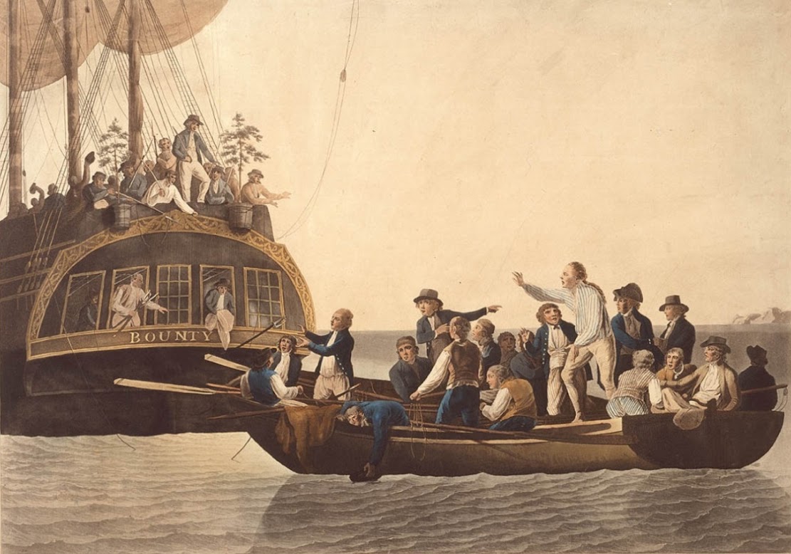
Polynesia: Forensic analysis of pigtails to help identify original 'mutineers of H.M.S. Bounty'
Forensics: Intricate animal and flower tattoos found on Egyptian mummy

Forensics: Single strain of plague bacteria sparked multiple historical and modern pandemics
