The Great London:
Genetics

  • Natural Heritage: Sampling species' DNA trails is leading to better environmental monitoring

    Natural Heritage: Sampling species' DNA trails is leading to better environmental monitoring

    Using a technique that can tell if a species has passed by from just a sample of water, scientists are developing new ways to assess ecosystems.

    Sampling species' DNA trails is leading to better environmental monitoring
    Great crested newt [Credit: Imperial College London]

    All animals shed fragments of DNA as they go about their lives – in faeces, mucous, sperm and eggs, shed skin, hair and, eventually, their carcasses.

    These traces of genetic material can persist in the environment for some time – a matter of weeks in water and up to a few centuries in soil. With new, more sensitive DNA amplification and sequencing techniques, scientists can collect and analyse these fragments in water and soil samples and identify individual species that have passed by.

    One area where environmental DNA, or eDNA, is finding practical use is in environmental assessments, for example to check whether any protected species are present before construction works are carried out. Already, Defra in the UK have approved the use of eDNA sampling to assess the presence of protected great crested newts in ponds.

    Now, in a new partnership between Imperial College London and environmental ecology consultancy Thomson Ecology, scientists are hoping to expand the use of eDNA. They want to create protocols to assess whether different areas are home to key protected species, including crayfish, water voles, otters and reptiles.

    As well as looking at key protected species for conservation, the team want to use eDNA for biosecurity, by identifying invasive species. For example, as well as native crayfish, some UK waters have been occupied by invasive American Signal Crayfish, which outcompete the native species and damage the local environment. Early detection of invasive crayfish could mean they are dealt with sooner, and cause less damage.

    Ultimately, the researchers hope to be able to use eDNA to profile entire ecosystems, analysing water samples to get a snapshot of all the organisms present in the local environment that have shed some DNA.

    Victoria Priestley, who is taking on this task for her PhD thesis in the Department of Life Sciences at Imperial, said: "I think eDNA surveys represent a sea change in how we approach survey and monitoring of species.

    "There is a lot of effort going into eDNA research globally and once it becomes more established, we should be able to assess what species are present in an area much more quickly. Ultimately we should be able to use it to create a clearer and more detailed picture of global biodiversity."

    Efficient Environmental Assessments

    Currently, species are assessed based on intensive field surveys, requiring taxonomic expertise and often involving tagging animals and repeat visits to a site. However, Professor Vincent Savolainen, from the Department of Life Sciences at Imperial, is developing new protocols for various species.

    This is paving the way for much simpler and more cost-effective surveying for environmental assessments. Professor Savolainen said: "This research will contribute to developing new indices to meet goals of the Intergovernmental Science-Policy Platform on Biodiversity and Ecosystem Services (IPBES), the body that assesses the state of biodiversity and of the ecosystem services it provides to society, in response to requests from decision makers."

    Although sequencing techniques have improved dramatically in the last few decades, challenges remain in analysing eDNA. The fragments degrade over time, a process enhanced by temperature, microbes, enzymes and salinity.

    The rate that eDNA is 'shed' from species to species and individual to individual also requires more research, as does the role of predators in moving eDNA between sites, and especially how eDNA is distributed in aquatic environments.

    However, Priestley is positive that eDNA surveys have a bright future: "There is still some way to go before whole-ecosystem eDNA monitoring is standard practice, but I believe that at least in the near future, eDNA will increasingly be one of the options in the survey toolkit, working alongside traditional methods to obtain the best ecological survey data in the most efficient way."

    Positive Partnership

    Professor Tom Welton, Dean of the Faculty of Natural Sciences, said partnerships like this one help translate research into real-world applications: "This exciting collaboration demonstrates that research across the whole breadth of natural sciences at Imperial, even on newts, has practical applications to real world problems.

    "Our partnership with Thomson Ecology will allow our research to have a positive impact on environmental protection and conservation."

    Author: Hayley Dunning | Source: Imperial College London [November 25, 2016]

  • Evolution: Sex cells evolved to pass on quality mitochondria

    Evolution: Sex cells evolved to pass on quality mitochondria

    Mammals immortalise their genes through eggs and sperm to ensure future generations inherit good quality mitochondria to power the body's cells, according to new UCL research.

    Sex cells evolved to pass on quality mitochondria
    One of a series of ova made in a spell of reproductive mitochondrial interest. The ovum about to ovulate has differentiated 
    from the rest of the surrounding tissue and is getting ready to leave the ovary. Its mitochondria are organized mainly 
    around the nucleus. The cell is full of potential and force. A big journey of life may be about to start 
    [Credit: Odra Noel]

    Before now, it was not known why mammals rely on dedicated sex cells that are formed early in development (a germline) to make offspring whereas plants and other simple animals, such as corals and sponges, use sex cells produced later in life from normal body tissues.

    In a new study, published today in >PLOS Biology and funded by Natural Environment Research Council, Engineering & Physical Sciences Research Council and the Leverhulme Trust, UCL scientists developed an evolutionary model to investigate how these differences evolved over time and discovered that the germline in mammals developed in response to selection on mitochondria (the powerhouses of cells).

    First author and UCL PhD student, Arunas Radzvilavicius, said: "There have been many theories about why mammals have a specialised germline when plants and other ancient animals don't. Some suggest it was due to complexity of tissues or a selfish conflict between cells. The distinction between sex cells and normal body tissues seems to be necessary for the evolution of very complex specialised tissues like brain.

    "Surprisingly, we found that these aren't the reason. Rather, it's about the number of genetic mutations in mitochondrial DNA over time, which differs between organisms, and the variation between cells caused by the mitochondria being randomly partitioned into daughter cells at each division."

    In plants, mitochondrial mutations creep in slowly, so a germline isn't needed as mutations are corrected by natural selection. Mitochondrial variation is maximised by forming the next generation from the same cells used to make normal tissue cells. When the cells divide to form new daughter cells, some receive more mutant mitochondria than others and these cells are then removed through natural selection, preserving the reproductive cells containing higher quality mitochondria.

    In mammals, genetic errors in mitochondria accumulate more quickly due to our higher metabolic rate so using cells that have undergone lots of division cycles would be a liability. Mitochondria are therefore only passed along to the next generation through a dedicated female germline in the form of large eggs. This protects against errors being introduced as eggs undergo many fewer replication cycles than cells in other tissues such as the gut, skin and blood.

    The germline ensures that the best quality mitochondria are transferred but restricts the genetic variation in the next generation of cells in the developing embryo. This is corrected for by mammals generating far too many egg cells which are removed during development. For example, humans are born with over 6 million egg-precursor cells, 90% of which are culled by the start of puberty in a mysterious process called atresia.

    Senior author, Dr Nick Lane (UCL CoMPLEX and Genetics, Evolution & Environment) added: "We think the rise in mitochondrial mutation rate likely occurred in the Cambrian explosion 550 million years ago when oxygen levels rose. This was the first appearance of motile animals in the fossil record, things like trilobites that had eyes and armour plating - predators and prey. By moving around they used their mitochondria more and that increased the mutation rate. So to avoid these mutations accumulating they needed to have fewer rounds of cell division, and that meant sequestering a specialized germline."

    Co-author, Professor Andrew Pomiankowski (UCL Genetics, Evolution & Environment), concluded: "Without a germline, animals with complex development and brains could not exist. Scientists have long tried to explain the evolution of the germline in terms of complexity. Who would have thought it arose from selection on mitochondrial genes? We hope our discovery will transform the way researchers understand animal development, reproduction and aging."

    Source: University College London [December 20, 2016]

  • Genetics: Obesity in humans linked to fat gene in prehistoric apes
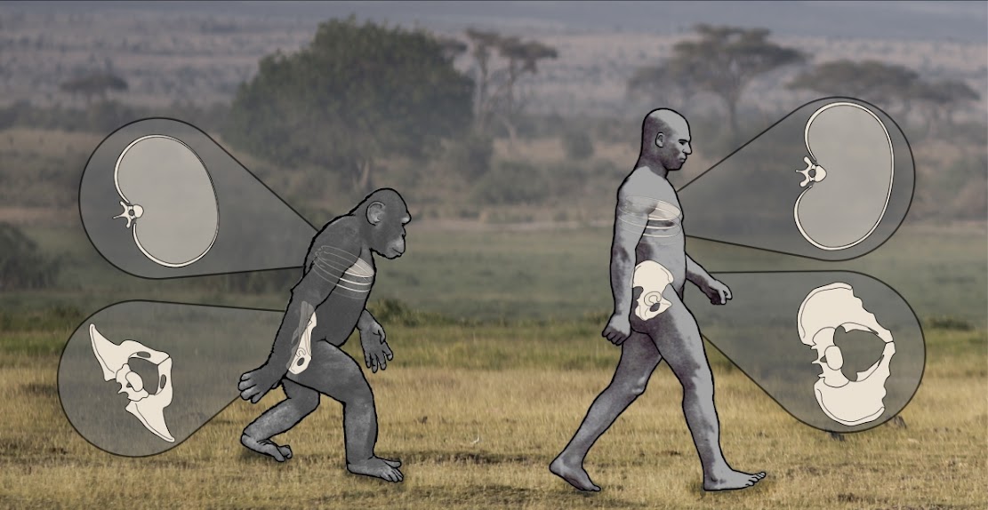
    Genetics: Obesity in humans linked to fat gene in prehistoric apes

    A genetic mutation in extinct European apes that enabled them to convert fruit sugar into fat could be a cause of the modern obesity epidemic and diabetes, according to scientists. Fossil evidence reveals that apes living around 16 million years ago, in what was then subtropical Europe, began to suffer as global cooling subsequently changed the forest, making the fruit they ate scarce.

    Obesity in humans linked to fat gene in prehistoric apes
    Extinct European apes evolved into today's great apes and the earliest hominids 
    [Credit: Nathan Thompson, Lucille Betti-Nash, and Deming Yang]

    Experts suggest that a mutation in the uricase gene which helps to convert fruit sugar (fructose) occurred around 15 million years ago. This aided apes in adding on fat layers so they could survive famines and harsh winters.

    Persistence of the same mutation in all modern great apes and all modern humans, along with the fossil evidence, suggests that the now extinct European apes evolved into today's great apes and the earliest hominids.

    Scientists have spent decades researching the genetic causes of obesity which is rarely found in other animals – apart from domesticated pets. The latest research focuses on fructose: a sugar which breaks down to form uric acid in the blood, according to a Sunday Times report.

    The Western diet contains so much uric acid that it cannot be removed quickly enough, but triggers liver cells to turn fructose into fat, with the effect of humans adding on extra weight. The uricase mutation predisposes humans to obesity and diabetes in modern times. The results suggest a need to eat and drink much less fructose to fight obesity and prevent its dangerous complications.

    "The gene enables uric acid levels to spike in response to two types of food," wrote Peter Andrews, professor of anthropology at University College London in Scientific American, co-authored with Richard Johnson, a professor of medicine in the US. "Those like beer that produce a lot of uric acid [directly] and those that contain a lot of fructose. These include honey and processed food that are high in table sugar or high-fructose corn syrup. When uric acid spikes we become susceptible to obesity and diabetes."

    Obesity is considered as one of the biggest public health challenges of the century. Statistics show that it is affecting more than 500 million people worldwide. In the US alone, obesity costs at least $200 billion each year.

    This medical condition also contributes to potentially fatal disorders such as cancer, type 2 diabetes and cardiovascular disease.

    Author: Fiona Keating | Source: International Business Times [November 20, 2015]

  • Genetics: Scientists sequence ancient British 'gladiator' genomes from Roman York

    Genetics: Scientists sequence ancient British 'gladiator' genomes from Roman York

    Cutting-edge genome technology in Trinity College Dublin has cast more light on a mystery that has perplexed archaeologists for more than a decade. The origins of a set of Roman-age decapitated bodies, found by York Archaeological Trust at Driffield Terrace in the city, have been explored, revealing a Middle Eastern body alongside native British.

    Scientists sequence ancient British 'gladiator' genomes from Roman York
    One of the skeletons excavated by York Archaeological Trust at Driffield Terrace
    [Credit: York Archaeological Trust]

    Archaeologists have speculated that the skeletons belonged to gladiators, although they could also have been soldiers or criminals. Several suffered perimortem decapitation and were all of a similar age – under 45 years old. Their skulls were buried with the body, although not positioned consistently – some were on the chest, some within the legs, and others at the feet.

    Although examining the skeletons revealed much about the life they lived – including childhood deprivation and injuries consistent with battle trauma – it was not until genomic analysis by a team from Trinity College Dublin, led by Professor of Population Genetics, Dan Bradley, that archaeologists could start to piece together the origins of the men.

    The Trinity College team recently published the first prehistoric Irish genomes and this analysis by Trinity PhD Researcher, Rui Martiniano, also breaks new ground as it represents the first genome analysis of ancient Britons.

    From the skeletons of more than 80 individuals, Dr Gundula Muldner of the University of Reading, Dr Janet Montgomery of the University of Durham and Malin Holst and Anwen Caffel of York Osteoarchaeology selected seven for whole genome analyses. Despite variation in isotope levels which suggested some of the 80 individuals lived their early lives outside Britain, most of those sampled had genomes similar to an earlier Iron Age woman from Melton, East Yorkshire. The poor childhood health of these men suggests that they were locals who endured childhood stress, but their robust skeletons and healed trauma, suggest that they were used to wielding weapons.

    Scientists sequence ancient British 'gladiator' genomes from Roman York
    The Roman-age skeletons from Driffield Terrace laid out in York's Guildhall 
    [Credit: York Archaeological Trust]

    The nearest modern descendants of the Roman British men sampled live not in Yorkshire, but in Wales. A man from a Christian Anglo-Saxon cemetery in the village of Norton, Teesside, has genes more closely aligned to modern East Anglia and Dutch individuals and highlights the impact of later migrations upon the genetic makeup of the earlier Roman British inhabitants.

    However, one of the decapitated Romans had a very different story, of Middle Eastern origin he grew up in the region of modern day Palestine, Jordan or Syria before migrating to this region and meeting his death in York.

    "Archaeology and osteoarchaeology can tell us a certain amount about the skeletons, but this new genomic and isotopic research can not only tell us about the body we see, but about its origins, and that is a huge step forward in understanding populations, migration patterns and how people moved around the ancient world," says Christine McDonnell, Head of Curatorial and Archive Services for York Archaeological Trust.

    "This hugely exciting, pioneering work will become the new standard for understanding the origins of skeletons in the future, and as the field grows, and costs of undertaking this kind of investigation fall, we may be able to refine our knowledge of exactly where the bodies were born to a much smaller region. That is a remarkable advance."

    Scientists sequence ancient British 'gladiator' genomes from Roman York
    The Roman skeletons were found at Driffield Terrace in York with their skulls placed between their legs,
     at their feet or on their chests [Credit: York Archaeological Trust]

    As well as Trinity College Dublin, the multi-disciplinary scientific analysis involved scientists from the University of York and The York Archaeological Trust, as well as the universities of Durham, Reading and Sheffield, University College London and the University Medical Centre in Utrecht. The research also included experts from York Osteoarchaeology Ltd, City of York Council and the Natural History Museum.

    The Roman skeletons sampled were all male, under 45 years old and most had evidence of decapitation. They were taller than average for Roman Britain and displayed evidence of significant trauma potentially related to interpersonal violence. All but one would have had brown eyes and black or brown hair but one had distinctive blue eyes and blond hair similar to the single Anglo-Saxon individual.

    The demographic profile of the York skeletons resembles the population structure in a Roman burial ground believed to be for gladiators at Ephesus. But the evidence could also fit with a military context—the Roman army had a minimum recruitment height and fallen soldiers would match the age profile of the York cemetery.

    Professor Dan Bradley, Trinity, said: "Whichever the identity of the enigmatic headless Romans from York, our sample of the genomes of seven of them, when combined with isotopic evidence, indicate six to be of British origin and one to have origins in the Middle East. It confirms the cosmopolitan character of the Roman Empire even at its most northerly extent."

    PhD Researcher and lead author, Rui Martiniano, Trinity, said: "This is the first refined genomic evidence for far-reaching ancient mobility and also the first snapshot of British genomes in the early centuries AD, indicating continuity with an Iron Age sample before the migrations of the Anglo-Saxon period."

    Professor Matthew Collins, of the BioArCh research facility in the Department of Archaeology at York, who co-ordinated the report on the research, "These genomes give the first snapshot of British genomes in the early centuries AD, showing continuity with the earlier Iron Age and evidence of migrations in the Anglo-Saxon period."

    The paper is published in >Nature Communications.

    Source: Trinity College Dublin [January 20, 2016]

  • Evolution: Rooting the family tree of placental mammals

    Evolution: Rooting the family tree of placental mammals

    Placental mammals consist of three main groups that diverged rapidly, evolving in wildly different directions: Afrotheria (for example, elephants and tenrecs), Xenarthra (such as armadillos and sloths) and Boreoeutheria (all other placental mammals). The relationships between them have been a subject of fierce controversy with multiple studies coming to incompatible conclusions over the last decade leading some researchers to suggest that these relationships might be impossible to resolve.

    Rooting the family tree of placental mammals
    Xenarthra, the group to which sloths such as this belong, is one of three main 
    groups of placental mammals that diverged rapidly, evolving in wildly
     different directions [Credit: University of Bristol]

    There are thus many outstanding questions such as which is the oldest sibling of the three? Did the mammals go their separate ways due to South America and Africa breaking apart? And if not, when did placentals split up?

    "This has been one of the areas of greatest debate in evolutionary biology, with many researchers considering it impossible to resolve," said lead author Dr Tarver of Bristol's School of Earth Sciences. "Now we've proven these problems can be solved -- you just need to analyse genome-scale datasets using models that accurately reflect genomic evolution."

    The researchers assembled the largest mammalian phylogenomic dataset ever collected before testing it with a variety of models of molecular evolution, choosing the most robust model and then analysing the data using several supercomputer clusters at the University of Bristol and the University of Texas Advanced Computing Centre. "We tested it to destruction," said Dr Tarver. "We threw the kitchen sink at it."

    "A complication in reconstructing evolutionary histories from genomic data is that different parts of genomes can and often do give conflicting accounts of the history," said Dr Siavash Mirarab at the University of California San Diego, USA. "Individual genes within the same species can have different histories. This is one reason why the controversy has stood so long -- many thought the relationships couldn't be resolved."

    To address the complexities of analysing large numbers of genes shared among many species, the researchers paired two fundamentally different approaches -- concatenated and coalescent-based analyses -- to confirm the findings. When the dust settled, the team had a specific family tree showing that Atlantogenata (containing the sibling groups of African Afrotheria and the South American Xenarthra) is the sister group to all other placentals.

    Because many conflicting family trees have already been published, the team then gathered three of the most influential rivals and tested them against each other with the same model. All of the previous studies suddenly fell into line, their data agreeing with Tarver and colleagues.

    With the origins of the family tree resolved, what does this mean for placental mammals? The researchers folded in another layer -- a molecular clock analysis. "The molecular clock analysis uses a combination of fossils and genomic data to estimate when these lineages diverged from each other," said author Dr Mario Dos-Reis of Queen Mary London, UK. "The results show that the afrotherians and xenarthrens diverged from one another around 90 million years ago."

    Previously, scientists thought that when Africa and South America separated from each other over 100 million years ago, they broke up the family of placental mammals, who went their separate evolutionary ways divided by geography. But the researchers found that placental mammals didn't split up until after Africa and South America had already separated.

    "We propose that South America's living endemic Xenarthra (for exmaple, sloths, anteaters, and armadillos) colonized the island-continent via overwater dispersal," said study author Dr Rob Asher of the University of Cambridge, UK.

    Dr Asher suggests that this isn't as difficult as you might think. Mammals are among the great adventurers of the animal kingdom, and at the time the proto-Atlantic was only a few hundred miles wide. We already know that New World monkeys crossed the Atlantic later, when it was much bigger, probably on rafts formed from storm debris. And, of course, mammals repeatedly colonised remote islands like Madagascar.

    "You don't always need to overturn the status quo to make a big impact," said Dr Tarver. "All of the competing hypotheses had some evidence to support them -- that's precisely why it was the source of such controversy. Proving the roots of the placental family tree with hard empirical evidence is a massive accomplishment."

    The findings are published in Genome Biology and Evolution journal.

    Source: University of Bristol [February 15, 2016]

  • Genetics: DNA analysis reveals Roman London was a multi-ethnic melting pot

    Genetics: DNA analysis reveals Roman London was a multi-ethnic melting pot

    A DNA analysis of four ancient Roman skeletons found in London shows the first inhabitants of the city were a multi-ethnic mix similar to contemporary Londoners, the Museum of London said on Monday.

    DNA analysis reveals Roman London was a multi-ethnic melting pot
    The displayed skeleton of "The Harper Road Woman", one of four 
    ancient Roman skeletons that have undergone DNA analysis 
    [Credit: Museum of London/AFP]

    Two of the skeletons were of people born outside Britain -- one of a man linked genealogically to eastern Europe and the Near East, the other of a teenage girl with blue eyes from north Africa.

    The injuries to the man's skull suggest that he may have been killed in the city's amphitheatre before his head was dumped into an open pit.

    Both the man and the girl were suffering from periodontal disease, a type of gum disease.

    The other two skeletons of people believed to have been born in Britain were of a woman with maternal ancestry from northern Europe and of a man also with links through his mother to Europe or north Africa.

    "We have always understood that Roman London was a culturally diverse place and now science is giving us certainty," said Caroline McDonald, senior curator of Roman London at the museum.

    "People born in Londinium lived alongside people from across the Roman Empire exchanging ideas and cultures, much like the London we know today," she said.

    The museum said in a statement that this was "the first multidisciplinary study of the inhabitants of a city anywhere in the Roman Empire".

    The Romans founded Britain's capital city in the middle of the first century AD, under the emperor Claudius.

    Britain's University of Durham researched stable isotopes from tooth enamel to determine migration patterns.

    A tooth from each skeleton was also sent to McMaster University in Canada for DNA analysis that established the hair and eye colour of each individual and identified the diseases they were suffering from.

    McMaster University also examined the mitochondrial DNA (mtDNA) to identify maternal ancestry.

    The exhibition of the four skeletons, entitled "Written in Bone", opens on Friday.

    Source: AFP [November 24, 2015]

  • Forensics: Slavery carried bilharzia parasites from West Africa to the Caribbean

    Forensics: Slavery carried bilharzia parasites from West Africa to the Caribbean

    Scientists used the full DNA sequences of Schistosoma mansoni parasites from Africa and the French Caribbean to discover the fluke's origins, map its historic transmission and identify the secrets of its success. Their findings, published in Scientific Reports, show how the global slave trade transported the disease from Senegal and Cameroon to Guadeloupe. Further genomic comparison with a closely related schistosome species that infects rodents reveals how the parasite has adapted to infecting human beings.

    Slavery carried bilharzia parasites from West Africa to the Caribbean
    The bilharzia-causing parasite, Schistosoma mansoni,  first infected humans as they fished
     in lakes in East Africa and was spread, first to West Africa and then to the New World, 
    by slave traders in 16th-19th Centuries, genomics reveals 
    [Credit: Wellcome Trust Sanger Institute]

    Schistosoma mansoni is a blood fluke (flatworm) that infects more than 250 million people worldwide and causes more than 11,000 deaths each year. Six years ago the Sanger Institute published the parasite's first full DNA sequence (genome); this latest study used that 'genetic map' to construct and compare the genomes of S. mansoni parasites gathered from across Africa and the New World, the majority of which were held at the Schistosomiasis Collection in the Natural History Museum, London.

    By analysing the differences between the human-infecting S. mansoni and its close relative, the rodent-infecting S. rodhaini, the scientists calculated that the two species evolved from a common ancestor approximately 107,000 to 148,000 years ago in East Africa. This finding suggests that the species is much 'younger' than previously thought.

    "The timing of the separation of the two species coincidences with the first archaeological evidence of fishing in Africa," explains Thomas Crellen, first author of the study from Imperial College London, the Sanger Institute and the Royal Veterinary College London. "The parasite develops in freshwater and infects people by burrowing through their skin. The introduction of fishing would have meant that people spent more time in the water, greatly increasing their chances of being infected."

    Analysing the differences between genomes from different locations also revealed the darker side of human history.

    "Comparing the S. mansoni genomes suggests that flukes in West Africa split from their Caribbean counterparts at some point between 1117AD and 1742AD, which overlaps with the time of the 16th-19th Century Atlantic Slave Trade," says Professor Joanne Webster from Imperial College London and the Royal Veterinary College. "During this period more than 22,000 African people were transported from West Africa to Guadeloupe by French slave ships, and the fluke was carried with them."

    Comparing the genomes of S. mansoni with S. rodhaini also revealed the genetic variations that have been positively selected over time in the human-infecting fluke and have been "fixed" into its DNA. It is likely that these variations are the evolutionary adaptations that have occurred to enable the fluke to successfully tunnel into, and thrive within, human beings.

    "When we looked for the differences between human-infecting S. mansoni DNA and its rodent infecting cousin S. rodhaini, we found two important variations. We found that changes to two genes in S. mansoni's DNA -- VAL21 and an elastase gene -appear to be important in allowing the fluke to enter and live in humans," says Dr James Cotton, senior author of the study from the Sanger Institute. "VAL genes produce proteins that cause allergic responses, so it is possible that the variation in VAL21 helps the fluke to hide from our immune systems. The elastase gene helps the parasite to burrow in to the body, by breaking down elastin -- a major component of human skin."

    It is hoped that exploring the genetic makeup of the fluke it will be possible to discover more about the processes the parasite relies on to infect humans and offer new opportunities to develop preventive and therapeutic interventions.

    Source: Wellcome Trust Sanger Institute [February 17, 2016]

  • Genetics: Mummies from Hungary reveal TB's Roman lineage

    Genetics: Mummies from Hungary reveal TB's Roman lineage

    Bodies found in a 200 year-old Hungarian crypt have revealed the secrets of how tuberculosis (TB) took hold in 18th century Europe, according to a research team led by the University of Warwick.

    Mummies from Hungary reveal TB's Roman lineage
    One of the 265 mummies resting in cardboard boxes in the Hungarian 
    Natural History Museum in Budapest, Hungary
    [Credit: AP/Bela Szandelszky]

    A new study published in Nature Communications details how samples taken from naturally mummified bodies found in an 18th century crypt in the Dominican church of Vác in Hungary have yielded 14 tuberculosis genomes, suggesting that mixed infections were common when TB was at peak prevalence in Europe.

    The research team included collaborators from the Universities of Warwick and Birmingham, University College London, the Hebrew University in Jerusalem and the Hungarian Natural History Museum in Budapest. Lead author Professor Mark Pallen, from Warwick Medical School, said the discovery was significant for current and future infection control and diagnosis.

    Professor Pallen said: “Microbiological analyses of samples from contemporary TB patients usually report a single strain of tuberculosis per patient. By contrast, five of the eight bodies in our study yielded more than one type of tuberculosis – remarkably from one individual we obtained evidence of three distinct strains.”

    The team used a technique called “metagenomics” to identify TB DNA in the historical specimens—that is direct sequencing of DNA from samples without growing bacteria or deliberately fishing out TB DNA. This approach draws on the remarkable throughput and ease of use of modern DNA sequencing technologies.

    Gemma Kay, first author on the paper says: “Poignantly, we found evidence of an intimate link between strains from in a middle-aged mother and her grown-up daughter, suggesting both family members died from this devastating infection.”

    The team used the 18th century sequences to date the origin of the lineage of TB strains commonly found in Europe and America to the late Roman period, which fits in with the recent controversial suggestion that the most recent common ancestor of all TB strains occurred as recently as six thousand years ago.

    Professor Pallen said: “By showing that historical strains can be accurately mapped to contemporary lineages, we have ruled out, for early modern Europe, the kind of scenario recently proposed for the Americas—that is wholesale replacement of one major lineage by another—and have confirmed the genotypic continuity of an infection that has ravaged the heart of Europe since prehistoric times.”

    Professor Pallen added that with TB resurgent in many parts of the world, the struggle to contain this ancient infection was far from over. He concludes: “We have shown that metagenomic approaches can document past infections. However, we have also recently shown that metagenomics can identify and characterize pathogens in contemporary samples, so such approaches might soon also inform current and future infectious disease diagnosis and control.”

    For more photos of the Hungarian mummies visit the website Morbid Anatomy.

    Source: University of Warwick [April 07, 2015]

  • Breaking News: Complex genetic ancestry of Americans uncovered

    Breaking News: Complex genetic ancestry of Americans uncovered

    By comparing the genes of current-day North and South Americans with African and European populations, an Oxford University study has found the genetic fingerprints of the slave trade and colonization that shaped migrations to the Americas hundreds of years ago.

    Complex genetic ancestry of Americans uncovered
    A 1770 painting showing Spanish, Peruvian and mixed-race people
    [Credit: WikiCommons]

    The study published in Nature Communications found that:

    • While Spaniards provide the majority of European ancestry in continental American Hispanic/Latino populations, the most common European genetic source in African-Americans and Barbadians comes from Great Britain.
    • The Basques, a distinct ethnic group spread across current-day Spain and France, provided a small but distinct genetic contribution to current-day Continental South American populations, including the Maya in Mexico.
    • The Caribbean Islands of Puerto Rico and the Dominican Republic are genetically similar to each other and distinct from the other populations, probably reflecting a different migration pattern between the Caribbean and mainland America.
    • Compared to South Americans, people from Caribbean countries (such as the Barbados) had a larger genetic contribution from Africa.
    • The ancestors of current-day Yoruba people from West Africa (one of the largest African ethnic groups) provided the largest contribution of genes from Africa to all current-day American populations.
    • The proportion of African ancestry varied across the continent, from virtually zero (in the Maya people from Mexico) to 87% in current-day Barbados.
    • South Italy and Sicily also provided a significant European genetic contribution to Colombia and Puerto Rico, in line with the known history of Italian emigrants to the Americas in the late 19th and early 20th century.
    • One of the African-American groups from the USA had French ancestry, in agreement with historical French immigration into the colonial Southern United States.
    • The proportion of genes from European versus African sources varied greatly from individual to individual within recipient populations.

    The team, which also included researchers from UCL (University College London) and the Universita' del Sacro Cuore of Rome, analysed more than 4,000 previously collected DNA samples from 64 different populations, covering multiple locations in Europe, Africa and the Americas. Since migration has generally flowed from Africa and Europe to the Americas over the last few hundred years, the team compared the 'donor' African and European populations with 'recipient' American populations to track where the ancestors of current-day North and South Americans came from.

    'We found that the genetic profile of Americans is much more complex than previously thought,' said study leader Professor Cristian Capelli from the Department of Zoology.

    The research team analysed DNA samples collected from people in Barbados, Columbia, the Dominican Republic, Ecuador, Mexico, Puerto Rico and African-Americans in the USA.

    They used a technique called haplotype-based analysis to compare the pattern of genes in these 'recipient populations' to 'donor populations' in areas where migrants to America came from.

    'We firstly grouped subsets of people in Africa and Europe who were genetically similar and used this fine scale resolution to find which combinations of these clusters resulted in the sort of mixtures that we now see in people across the Americas', said the study's first author, Dr Francesco Montinaro from the Department of Zoology.

    'We can see the huge genetic impact that the slave trade had on American populations and our data match historical records', said study author Dr Garrett Hellenthal from the UCL Genetics Institute, 'The majority of African Americans have ancestry similar to the Yoruba people in West Africa, confirming that most African slaves came from this region. In areas of the Americas historically under Spanish rule, populations also have ancestry related to what is now Senegal and Gambia. Records show that around a third of the slaves sent to Spanish America in the 17th Century came from this region, and we can see the genetic evidence of this in modern Americans really clearly.'

    These genetic findings also uncover previously unknown migration. ‘We found a clear genetic contribution from the Basques in modern-day Maya in Mexico’, said Professor Capelli. ‘This suggests that the Basque also took part in the colonisation of the Americas, coming over either with the Spanish conquistadores or in later waves of migration’.

    'The differences in European ancestry between the Caribbean islands and mainland American population that we found were also previously unknown. It is likely that these differences reflect different patterns of migration between the Caribbean and mainland America.'

    'These results show just how powerful a genetic approach can be when it comes to uncovering hidden patterns of ancestry. We hope to use the same approach to look at other populations with diverse genetic contributions, such as Brazilians,' said Professor Capelli.

    Source: University of Oxford [March 24, 2015]

  • Genetics: Genes for nose shape found

    Genetics: Genes for nose shape found

    Genes that drive the shape of human noses have been identified by a UCL-led study. The four genes mainly affect the width and 'pointiness' of noses which vary greatly between different populations. The new information adds to our understanding of how the human face evolved and may help contribute to forensic DNA technologies that build visual profiles based on an individual's genetic makeup.

    Genes for nose shape found
    Variation between nose shape and the specific genes responsible 
    [Credit: Dr Kaustubh Adhikari, UCL]

    The study, published today in >Nature Communications, analysed a population of over 6,000 people with varied ancestry across Latin America to study the differences in normal facial features and identify the genes which control the shape of the nose and chin.

    The researchers identified five genes which play a role in controlling the shape of specific facial features. DCHS2, RUNX2, GLI3 and PAX1 affect the width and pointiness of the nose and another gene -- EDAR -- affects chin protrusion.

    "Few studies have looked at how normal facial features develop and those that have only looked at European populations, which show less diversity than the group we studied. What we've found are specific genes which influence the shape and size of individual features, which hasn't been seen before.

    "Finding out the role each gene plays helps us to piece together the evolutionary path from Neanderthal to modern humans. It brings us closer to understanding how genes influence the way we look, which is important for forensics applications," said the first author of the report, Dr Kaustubh Adhikari, UCL Cell & Developmental Biology.

    People have different shaped facial features based on their genetic heritage and this is partly due to how the environment influenced the evolution of the human genome. The nose, for example, is important for regulating the temperature and humidity of the air we breathe in so developed different shapes in warmer and cooler climates.

    "It has long been speculated that the shape of the nose reflects the environment in which humans evolved. For example, the comparatively narrower nose of Europeans has been proposed to represent an adaptation to a cold, dry climate. Identifying genes affecting nose shape provides us with new tools to examine this question, as well as the evolution of the face in other species. It may also help us understand what goes wrong in genetic disorders involving facial abnormalities," explained Professor Andrés Ruiz-Linares UCL Biosciences, who led the study.

    The team collected and analysed DNA samples from 6,630 volunteers from the CANDELA cohort recruited in Brazil, Colombia, Chile, Mexico and Peru. After an initial screen, a sample size of 5,958 was used. This group included individuals of mixed European (50%), Native American (45%) and African (5%) ancestry, resulting in a large variation in facial features.

    Both men and women were assessed for 14 different facial features and whole genome analysis identified the genes driving differences in appearance.

    A subgroup of 3,000 individuals had their features assessed using a 3D reconstruction of the face in order to obtain exact measurements of facial features and the results identified the same genes.

    The study identified genes that are involved in bone and cartilage growth and the development of the face. GLI3, DCHS2 and PAX1 are all genes known to drive cartilage growth -- GLI3 gave the strongest signal for controlling the breadth of nostrils, DCHS2 was found to control nose 'pointiness' and PAX1 also influences nostril breadth. RUNX2 which drives bone growth was seen to control nose bridge width.

    The genes GLI3, DCHS2 and RUNX2 are known to show strong signals of recent selection in modern humans compared to archaic humans such as Neanderthals and Denisovans; GLI3 in particular undergoing rapid evolution.

    Source: University College London [May 19, 2016]

  • Genetics: First fine-scale genetic map of the British Isles
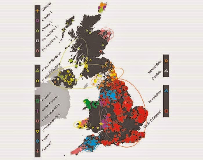
    Genetics: First fine-scale genetic map of the British Isles

    Many people in the UK feel a strong sense of regional identity, and it now appears that there may be a scientific basis to this feeling, according to a landmark new study into the genetic makeup of the British Isles.

    First fine-scale genetic map of the British Isles
    Subtle differences in the genes of more than 2,000 people in England, Scotland, Wales 
    and Northern Ireland reveal 17 distinct groups, represented by different symbols 
    and colors on the map [Credit: Stephen Leslie; Contains OS data 
    © Crown copyright and database right 2012; © EuroGeographics
     for some administrative boundaries]

    An international team, led by researchers from the University of Oxford, UCL (University College London) and the Murdoch Childrens Research Institute in Australia, used DNA samples collected from more than 2,000 people to create the first fine-scale genetic map of any country in the world.

    Their findings, published in Nature, show that prior to the mass migrations of the 20th century there was a striking pattern of rich but subtle genetic variation across the UK, with distinct groups of genetically similar individuals clustered together geographically.

    By comparing this information with DNA samples from over 6,000 Europeans, the team was also able to identify clear traces of the population movements into the UK over the past 10,000 years. Their work confirmed, and in many cases shed further light on, known historical migration patterns.

    Key findings

    • There was not a single "Celtic" genetic group. In fact the Celtic parts of the UK (Scotland, Northern Ireland, Wales and Cornwall) are among the most different from each other genetically. For example, the Cornish are much more similar genetically to other English groups than they are to the Welsh or the Scots.
    • There are separate genetic groups in Cornwall and Devon, with a division almost exactly along the modern county boundary.
    • The majority of eastern, central and southern England is made up of a single, relatively homogeneous, genetic group with a significant DNA contribution from Anglo-Saxon migrations (10-40% of total ancestry). This settles a historical controversy in showing that the Anglo-Saxons intermarried with, rather than replaced, the existing populations.
    • The population in Orkney emerged as the most genetically distinct, with 25% of DNA coming from Norwegian ancestors. This shows clearly that the Norse Viking invasion (9th century) did not simply replace the indigenous Orkney population.
    • The Welsh appear more similar to the earliest settlers of Britain after the last ice age than do other people in the UK.
    • There is no obvious genetic signature of the Danish Vikings, who controlled large parts of England ("The Danelaw") from the 9th century.
    • There is genetic evidence of the effect of the Landsker line -- the boundary between English-speaking people in south-west Pembrokeshire (sometimes known as "Little England beyond Wales") and the Welsh speakers in the rest of Wales, which persisted for almost a millennium.
    • The analyses suggest there was a substantial migration across the channel after the original post-ice-age settlers, but before Roman times. DNA from these migrants spread across England, Scotland, and Northern Ireland, but had little impact in Wales.
    • Many of the genetic clusters show similar locations to the tribal groupings and kingdoms around end of the 6th century, after the settlement of the Anglo-Saxons, suggesting these tribes and kingdoms may have maintained a regional identity for many centuries.

    The Wellcome Trust-funded People of the British Isles study analysed the DNA of 2,039 people from rural areas of the UK, whose four grandparents were all born within 80km of each other. Because a quarter of our genome comes from each of our grandparents, the researchers were effectively sampling DNA from these ancestors, allowing a snapshot of UK genetics in the late 19th Century. They also analysed data from 6,209 individuals from 10 (modern) European countries.

    To uncover the extremely subtle genetic differences among these individuals the researchers used cutting-edge statistical techniques, developed by four of the team members. They applied these methods, called fineSTRUCTURE and GLOBETROTTER, to analyse DNA differences at over 500,000 positions within the genome. They then separated the samples into genetically similar individuals, without knowing where in the UK the samples came from. By plotting each person onto a map of the British Isles, using the centre point of their grandparents' birth places, they were able to see how this distribution correlated with their genetic groupings.

    The researchers were then able to "zoom in" to examine the genetic patterns in the UK at levels of increasing resolution. At the broadest scale, the population in Orkney (islands to the north of Scotland) emerged as the most genetically distinct. At the next level, Wales forms a distinct genetic group, followed by a further division between north and south Wales. Then the north of England, Scotland, and Northern Ireland collectively separate from southern England, before Cornwall forms a separate cluster. Scotland and Northern Ireland then separate from northern England. The study eventually focused at the level where the UK was divided into 17 genetically distinct clusters of people.

    Dr Michael Dunn, Head of Genetics & Molecular Sciences at the Wellcome Trust, said: "These researchers have been able to use modern genetic techniques to provide answers to the centuries' old question -- where we come from. Beyond the fascinating insights into our history, this information could prove very useful from a health perspective, as building a picture of population genetics at this scale may in future help us to design better genetic studies to investigate disease."

    Source: Wellcome Trust [March 18, 2015]

  1. A treasure horde of art and history under one roof
  2. Art Institute announces major long-term loan of ancient Near Eastern statuette
  3. Treasure trove of medieval manuscripts published
  4. Tomb of the Scipios reopens to public in Rome
  5. Mexican city to begin countdown for Dec. 21, 2012